 Movie
Movie Controller

[English] 日本語
 Yorodumi
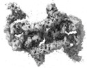
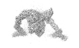
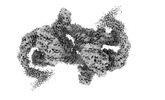
- EMDB-36134: Structure of CRL2APPBP2 bound with the C-degron of MRPL28 (dimer) -
+ Open data

- Basic information
| Entry |  | |||||||||
|---|---|---|---|---|---|---|---|---|---|---|
| Title | Structure of CRL2APPBP2 bound with the C-degron of MRPL28 (dimer) | |||||||||
 Map data Map data | ||||||||||
 Sample Sample |
| |||||||||
 Keywords Keywords | E3 Ubiquitination ligase /  PROTEIN BINDING PROTEIN BINDING | |||||||||
| Function / homology |  Function and homology information Function and homology informationubiquitin-dependent protein catabolic process via the C-end degron rule pathway / target-directed miRNA degradation / elongin complex / VCB complex / Cul5-RING ubiquitin ligase complex / microtubule associated complex / SCF-dependent proteasomal ubiquitin-dependent protein catabolic process / Cul2-RING ubiquitin ligase complex / SCF ubiquitin ligase complex / microtubule motor activity ...ubiquitin-dependent protein catabolic process via the C-end degron rule pathway / target-directed miRNA degradation / elongin complex / VCB complex / Cul5-RING ubiquitin ligase complex / microtubule associated complex / SCF-dependent proteasomal ubiquitin-dependent protein catabolic process / Cul2-RING ubiquitin ligase complex / SCF ubiquitin ligase complex / microtubule motor activity / ubiquitin ligase complex scaffold activity / Pausing and recovery of Tat-mediated HIV elongation / Tat-mediated HIV elongation arrest and recovery / HIV elongation arrest and recovery / Pausing and recovery of HIV elongation / ubiquitin-like ligase-substrate adaptor activity / intracellular transport / Tat-mediated elongation of the HIV-1 transcript / Formation of HIV-1 elongation complex containing HIV-1 Tat / Formation of HIV elongation complex in the absence of HIV Tat / RNA Polymerase II Transcription Elongation / Formation of RNA Pol II elongation complex / RNA Polymerase II Pre-transcription Events / intrinsic apoptotic signaling pathway / transcription corepressor binding / transcription elongation by RNA polymerase II / transcription initiation at RNA polymerase II promoter / TP53 Regulates Transcription of DNA Repair Genes / Vif-mediated degradation of APOBEC3G / intracellular protein transport / G1/S transition of mitotic cell cycle / cytoplasmic vesicle membrane / Oxygen-dependent proline hydroxylation of Hypoxia-inducible Factor Alpha / Inactivation of CSF3 (G-CSF) signaling / Regulation of expression of SLITs and ROBOs / ubiquitin-protein transferase activity / protein-macromolecule adaptor activity / positive regulation of proteasomal ubiquitin-dependent protein catabolic process / Antigen processing: Ubiquitination & Proteasome degradation / Neddylation / ubiquitin-dependent protein catabolic process / proteasome-mediated ubiquitin-dependent protein catabolic process / protein-containing complex assembly / microtubule / protein ubiquitination / ubiquitin protein ligase binding / protein-containing complex binding / nucleolus / regulation of transcription by RNA polymerase II / nucleoplasm / nucleus / cytosol / cytoplasmSimilarity search - Function | |||||||||
| Biological species |  Homo sapiens (human) Homo sapiens (human) | |||||||||
| Method | single particle reconstruction / cryo EM / Resolution: 3.22 Å | |||||||||
 Authors Authors | Zhao S / Zhang K / Xu C | |||||||||
| Funding support |  China, 1 items China, 1 items
| |||||||||
 Citation Citation | Journal: Proc Natl Acad Sci U S A / Year: 2023 Title: Molecular basis for C-degron recognition by CRL2 ubiquitin ligase. Authors: Shidong Zhao / Diana Olmayev-Yaakobov / Wenwen Ru / Shanshan Li / Xinyan Chen / Jiahai Zhang / Xuebiao Yao / Itay Koren / Kaiming Zhang / Chao Xu /  Abstract: E3 ubiquitin ligases determine the specificity of eukaryotic protein degradation by selective binding to destabilizing protein motifs, termed degrons, in substrates for ubiquitin-mediated proteolysis. ...E3 ubiquitin ligases determine the specificity of eukaryotic protein degradation by selective binding to destabilizing protein motifs, termed degrons, in substrates for ubiquitin-mediated proteolysis. The exposed C-terminal residues of proteins can act as C-degrons that are recognized by distinct substrate receptors (SRs) as part of dedicated cullin-RING E3 ubiquitin ligase (CRL) complexes. APPBP2, an SR of Cullin 2-RING ligase (CRL2), has been shown to recognize R-x-x-G/C-degron; however, the molecular mechanism of recognition remains elusive. By solving several cryogenic electron microscopy structures of active CRL2 bound with different R-x-x-G/C-degrons, we unveiled the molecular mechanisms underlying the assembly of the CRL2 dimer and tetramer, as well as C-degron recognition. The structural study, complemented by binding experiments and cell-based assays, demonstrates that APPBP2 specifically recognizes the R-x-x-G/C-degron via a bipartite mechanism; arginine and glycine, which play critical roles in C-degron recognition, accommodate distinct pockets that are spaced by two residues. In addition, the binding pocket is deep enough to enable the interaction of APPBP2 with the motif placed at or up to three residues upstream of the C-end. Overall, our study not only provides structural insight into CRL2-mediated protein turnover but also serves as the basis for future structure-based chemical probe design. | |||||||||
| History |
|
- Structure visualization
| Supplemental images |
|---|

- Downloads & links
-EMDB archive
| Map data | emd_36134.map.gz | 483.7 MB | EMDB map data format | |
|---|---|---|---|---|
| Header (meta data) | emd-36134-v30.xmlemd-36134.xml | 19 KB 19 KB | Display Display | EMDB header |
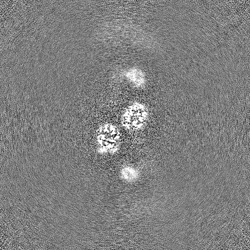
| Images |  emd_36134.png emd_36134.png | 49.2 KB | ||
| Filedesc metadata | emd-36134.cif.gz | 6.5 KB | ||
| Others | emd_36134_half_map_1.map.gzemd_36134_half_map_2.map.gz | 475.7 MB 475.7 MB | ||
| Archive directory |  http://ftp.pdbj.org/pub/emdb/structures/EMD-36134ftp://ftp.pdbj.org/pub/emdb/structures/EMD-36134 http://ftp.pdbj.org/pub/emdb/structures/EMD-36134ftp://ftp.pdbj.org/pub/emdb/structures/EMD-36134 | HTTPS FTP |
-Related structure data
| Related structure data |  8jauMC  8jalC 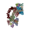 8jaqC  8jarC 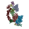 8jasC 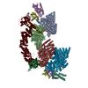 8javC M: atomic model generated by this map C: citing same article ( |
|---|---|
| Similar structure data |


-Links
| EMDB pages | EMDB (EBI/PDBe) / EMDataResource |
|---|---|
| Related items in Molecule of the Month |


-Map
| File | Download / File: emd_36134.map.gz / Format: CCP4 / Size: 512 MB / Type: IMAGE STORED AS FLOATING POINT NUMBER (4 BYTES) | ||||||||||||||||||||
|---|---|---|---|---|---|---|---|---|---|---|---|---|---|---|---|---|---|---|---|---|---|
| Voxel size | X=Y=Z: 0.82 Å | ||||||||||||||||||||
| Density |
| ||||||||||||||||||||
| Symmetry | Space group: 1 | ||||||||||||||||||||
| Details | EMDB XML:
|
-Supplemental data
-Half map: #1
| File | emd_36134_half_map_1.map | ||||||||||||
|---|---|---|---|---|---|---|---|---|---|---|---|---|---|
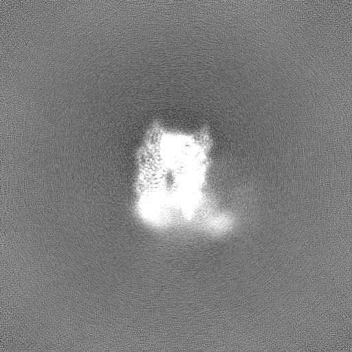
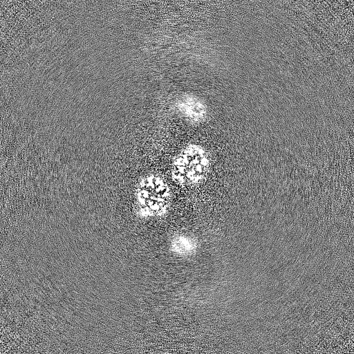
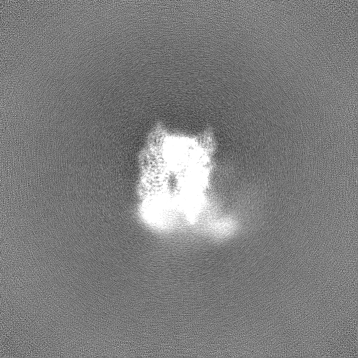
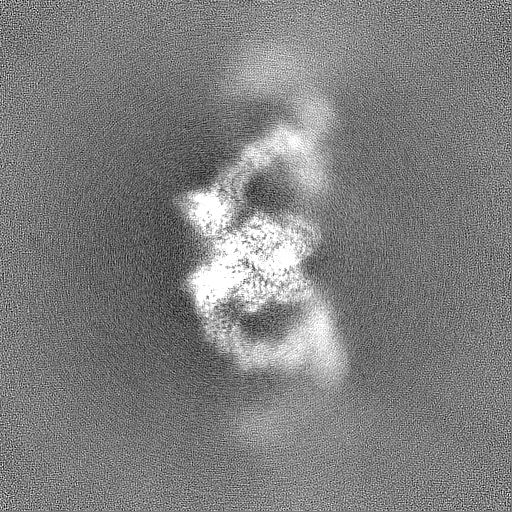
| Projections & Slices |
| ||||||||||||
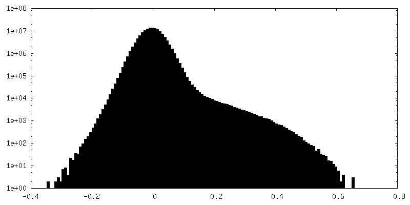
| Density Histograms |
 Z
Z Y
Y X
X


-Half map: #2
| File | emd_36134_half_map_2.map | ||||||||||||
|---|---|---|---|---|---|---|---|---|---|---|---|---|---|
| Projections & Slices |
| ||||||||||||
| Density Histograms |


- Sample components
-Entire : CRL2APPBP2 E3 liganse
| Entire | Name: CRL2APPBP2 E3 liganse |
|---|---|
| Components |
|
-Supramolecule #1: CRL2APPBP2 E3 liganse
| Supramolecule | Name: CRL2APPBP2 E3 liganse / type: complex / ID: 1 / Parent: 0 / Macromolecule list: #1-#5 |
|---|---|
| Source (natural) | Organism: Homo sapiens (human) |
| Molecular weight | Theoretical: 400 kDa/nm |
-Macromolecule #1: Amyloid protein-binding protein 2
| Macromolecule | Name: Amyloid protein-binding protein 2 / type: protein_or_peptide / ID: 1 / Number of copies: 2 / Enantiomer: LEVO |
|---|---|
| Source (natural) | Organism: Homo sapiens (human) |
| Molecular weight | Theoretical: 66.945258 KDa |
| Recombinant expression | Organism:  Escherichia coli (E. coli) Escherichia coli (E. coli) |
| Sequence | String: MAAVELEWIP ETLYNTAISA VVDNYIRSRR DIRSLPENIQ FDVYYKLYQQ GRLCQLGSEF CELEVFAKVL RALDKRHLLH HCFQALMDH GVKVASVLAY SFSRRCSYIA ESDAAVKEKA IQVGFVLGGF LSDAGWYSDA EKVFLSCLQL CTLHDEMLHW F RAVECCVR ...String: MAAVELEWIP ETLYNTAISA VVDNYIRSRR DIRSLPENIQ FDVYYKLYQQ GRLCQLGSEF CELEVFAKVL RALDKRHLLH HCFQALMDH GVKVASVLAY SFSRRCSYIA ESDAAVKEKA IQVGFVLGGF LSDAGWYSDA EKVFLSCLQL CTLHDEMLHW F RAVECCVR LLHVRNGNCK YHLGEETFKL AQTYMDKLSK HGQQANKAAL YGELCALLFA KSHYDEAYKW CIEAMKEITA GL PVKVVVD VLRQASKACV VKREFKKAEQ LIKHAVYLAR DHFGSKHPKY SDTLLDYGFY LLNVDNICQS VAIYQAALDI RQS VFGGKN IHVATAHEDL AYSSYVHQYS SGKFDNALFH AERAIGIITH ILPEDHLLLA SSKRVKALIL EEIAIDCHNK ETEQ RLLQE AHDLHLSSLQ LAKKAFGEFN VQTAKHYGNL GRLYQSMRKF KEAEEMHIKA IQIKEQLLGQ EDYEVALSVG HLASL YNYD MNQYENAEKL YLRSIAIGKK LFGEGYSGLE YDYRGLIKLY NSIGNYEKVF EYHNVLSNWN RLRDRQYSVT DALEDV STS PQSTEEVVQS FLISQNVEGP SC UniProtKB: Amyloid protein-binding protein 2 |
-Macromolecule #2: Cullin-2
| Macromolecule | Name: Cullin-2 / type: protein_or_peptide / ID: 2 / Number of copies: 2 / Enantiomer: LEVO |
|---|---|
| Source (natural) | Organism: Homo sapiens (human) |
| Molecular weight | Theoretical: 87.068836 KDa |
| Recombinant expression | Organism: Escherichia coli (E. coli) |
| Sequence | String: TSLKPRVVDF DETWNKLLTT IKAVVMLEYV ERATWNDRFS DIYALCVAYP EPLGERLYTE TKIFLENHVR HLHKRVLESE EQVLVMYHR YWEEYSKGAD YMDCLYRYLN TQFIKKNKLT EADLQYGYGG VDMNEPLMEI GELALDMWRK LMVEPLQAIL I RMLLREIK ...String: TSLKPRVVDF DETWNKLLTT IKAVVMLEYV ERATWNDRFS DIYALCVAYP EPLGERLYTE TKIFLENHVR HLHKRVLESE EQVLVMYHR YWEEYSKGAD YMDCLYRYLN TQFIKKNKLT EADLQYGYGG VDMNEPLMEI GELALDMWRK LMVEPLQAIL I RMLLREIK NDRGGEDPNQ KVIHGVINSF VHVEQYKKKF PLKFYQEIFE SPFLTETGEY YKQEASNLLQ ESNCSQYMEK VL GRLKDEE IRCRKYLHPS SYTKVIHECQ QRMVADHLQF LHAECHNIIR QEKKNDMANM YVLLRAVSTG LPHMIQELQN HIH DEGLRA TSNLTQENMP TLFVESVLEV HGKFVQLINT VLNGDQHFMS ALDKALTSVV NYREPKSVCK APELLAKYCD NLLK KSAKG MTENEVEDRL TSFITVFKYI DDKDVFQKFY ARMLAKRLIH GLSMSMDSEE AMINKLKQAC GYEFTSKLHR MYTDM SVSA DLNNKFNNFI KNQDTVIDLG ISFQIYVLQA GAWPLTQAPS STFAIPQELE KSVQMFELFY SQHFSGRKLT WLHYLC TGE VKMNYLGKPY VAMVTTYQMA VLLAFNNSET VSYKELQDST QMNEKELTKT IKSLLDVKMI NHDSEKEDID AESSFSL NM NFSSKRTKFK ITTSMQKDTP QEMEQTRSAV DEDRKMYLQA AIVRIMKARK VLRHNALIQE VISQSRARFN PSISMIKK C IEVLIDKQYI ERSQASADEY SYVA UniProtKB: Cullin-2 |
-Macromolecule #3: Elongin-B
| Macromolecule | Name: Elongin-B / type: protein_or_peptide / ID: 3 / Number of copies: 2 / Enantiomer: LEVO |
|---|---|
| Source (natural) | Organism: Homo sapiens (human) |
| Molecular weight | Theoretical: 13.147781 KDa |
| Recombinant expression | Organism: Escherichia coli (E. coli) |
| Sequence | String: MDVFLMIRRH KTTIFTDAKE SSTVFELKRI VEGILKRPPD EQRLYKDDQL LDDGKTLGEC GFTSQTARPQ APATVGLAFR ADDTFEALC IEPFSSPPEL PDVMKPQDSG SSANEQAVQ UniProtKB: Elongin-B |
-Macromolecule #4: Elongin-C
| Macromolecule | Name: Elongin-C / type: protein_or_peptide / ID: 4 / Number of copies: 2 / Enantiomer: LEVO |
|---|---|
| Source (natural) | Organism: Homo sapiens (human) |
| Molecular weight | Theoretical: 10.84342 KDa |
| Recombinant expression | Organism: Escherichia coli (E. coli) |
| Sequence | String: MYVKLISSDG HEFIVKREHA LTSGTIKAML SGPGQFAENE TNEVNFREIP SHVLSKVCMY FTYKVRYTNS STEIPEFPIA PEIALELLM AANFLDC UniProtKB: Elongin-C |
-Macromolecule #5: MRPL28 C-degron
| Macromolecule | Name: MRPL28 C-degron / type: protein_or_peptide / ID: 5 / Number of copies: 2 / Enantiomer: LEVO |
|---|---|
| Source (natural) | Organism: Homo sapiens (human) |
| Molecular weight | Theoretical: 1.670864 KDa |
| Recombinant expression | Organism: Escherichia coli (E. coli) |
| Sequence | String: QALSEPAVVQ KRASGQ |
-Macromolecule #6: ZINC ION
| Macromolecule | Name: ZINC ION / type: ligand / ID: 6 / Number of copies: 1 / Formula: ZN |
|---|---|
| Molecular weight | Theoretical: 65.409 Da |
-Experimental details
-Structure determination
| Method | cryo EM |
|---|---|
 Processing Processing | single particle reconstruction |
| Aggregation state | particle |
-Sample preparation
| Buffer | pH: 7.5 |
|---|---|
| Vitrification | Cryogen name: ETHANE |
- Electron microscopy
| Microscope | FEI TITAN KRIOS |
|---|---|
| Electron beam | Acceleration voltage: 300 kV / Electron source: FIELD EMISSION GUN |
| Electron optics | Illumination mode: OTHER / Imaging mode: BRIGHT FIELDBright-field microscopy / Nominal defocus max: 2.0 µm / Nominal defocus min: 0.8 µm |
| Image recording | Film or detector model: GATAN K3 (6k x 4k) / Average electron dose: 50.0 e/Å2 |
| Experimental equipment |  Model: Titan Krios / Image courtesy: FEI Company |
-Image processing
| Startup model | Type of model: OTHER / Details: Model from alphafold |
|---|---|
| Initial angle assignment | Type: ANGULAR RECONSTITUTION |
| Final angle assignment | Type: ANGULAR RECONSTITUTION |
| Final reconstruction | Resolution.type: BY AUTHOR / Resolution: 3.22 Å / Resolution method: FSC 0.143 CUT-OFF / Number images used: 156258 |
