 Movie
Movie Controller


+ Open data

- Basic information
| Entry |  | |||||||||
|---|---|---|---|---|---|---|---|---|---|---|
| Title | Cryo-EM structure of DIP-2I8I polymorph 2 | |||||||||
 Map data Map data | ||||||||||
 Sample Sample |
| |||||||||
 Keywords Keywords | Iodinated peptide / PROTEIN FIBRIL | |||||||||
| Biological species |   Homo sapiens (human) Homo sapiens (human) | |||||||||
| Method | helical reconstruction / cryo EM / Resolution: 3.75 Å | |||||||||
 Authors Authors | Li DN / Ma YY / Li D / Dai B / Liu C | |||||||||
| Funding support | 1 items
| |||||||||
 Citation Citation | Journal: To Be Published Title: Cryo-EM structure of DIP-2I8I fibril polymorph 2 Authors: Li DN / Ma YY / Li D / Dai B / Liu C | |||||||||
| History |
|
- Structure visualization
| Supplemental images |
|---|

- Downloads & links
-EMDB archive
| Map data | emd_35878.map.gz | 26.7 MB |  EMDB map data format EMDB map data format | |
|---|---|---|---|---|
| Header (meta data) | emd-35878-v30.xmlemd-35878.xml | 12 KB 12 KB | Display Display | EMDB header |
| FSC (resolution estimation) | emd_35878_fsc.xml | 14.8 KB | Display | FSC data file |
| Images | 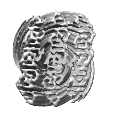 emd_35878.png emd_35878.png | 114.7 KB | ||
| Filedesc metadata | emd-35878.cif.gz | 4.2 KB | ||
| Others | emd_35878_half_map_1.map.gzemd_35878_half_map_2.map.gz | 218.4 MB 218.4 MB | ||
| Archive directory |  http://ftp.pdbj.org/pub/emdb/structures/EMD-35878ftp://ftp.pdbj.org/pub/emdb/structures/EMD-35878 http://ftp.pdbj.org/pub/emdb/structures/EMD-35878ftp://ftp.pdbj.org/pub/emdb/structures/EMD-35878 | HTTPS FTP |
-Related structure data

-Links
| EMDB pages | EMDB (EBI/PDBe) / EMDataResource |
|---|
-Map
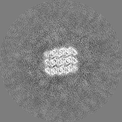
| File | Download / File: emd_35878.map.gz / Format: CCP4 / Size: 274.6 MB / Type: IMAGE STORED AS FLOATING POINT NUMBER (4 BYTES) | ||||||||||||||||||||||||||||||||||||
|---|---|---|---|---|---|---|---|---|---|---|---|---|---|---|---|---|---|---|---|---|---|---|---|---|---|---|---|---|---|---|---|---|---|---|---|---|---|
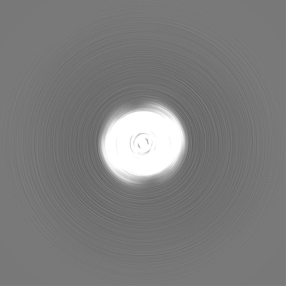
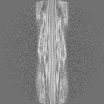
| Projections & slices | Image control
Images are generated by Spider. | ||||||||||||||||||||||||||||||||||||
| Voxel size | X=Y=Z: 0.83 Å | ||||||||||||||||||||||||||||||||||||
| Density |
| ||||||||||||||||||||||||||||||||||||
| Symmetry | Space group: 1 | ||||||||||||||||||||||||||||||||||||
| Details | EMDB XML:
|
 Z (Sec.)
 Y (Row.)
 X (Col.)


-Supplemental data
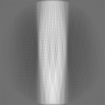
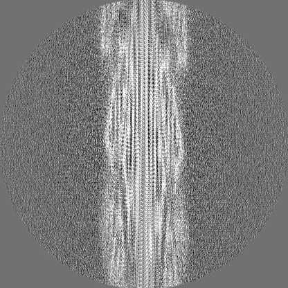
-Half map: #2
| File | emd_35878_half_map_1.map | ||||||||||||
|---|---|---|---|---|---|---|---|---|---|---|---|---|---|
| Projections & Slices |
| ||||||||||||
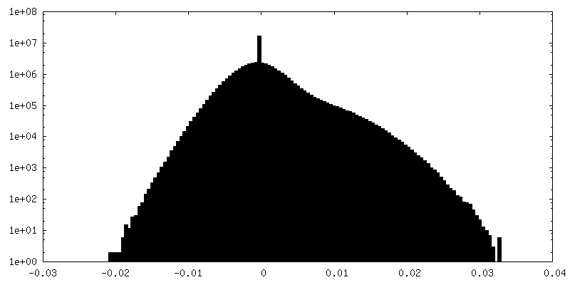
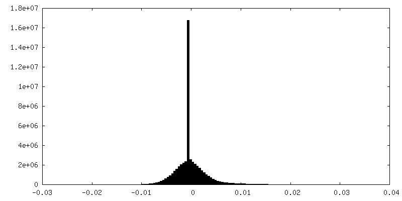
| Density Histograms |
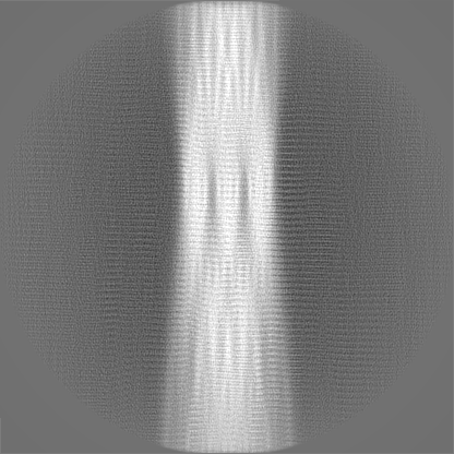
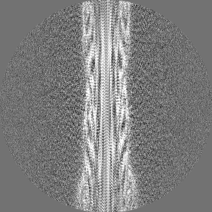

-Half map: #1
| File | emd_35878_half_map_2.map | ||||||||||||
|---|---|---|---|---|---|---|---|---|---|---|---|---|---|
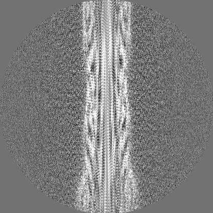
| Projections & Slices |
| ||||||||||||
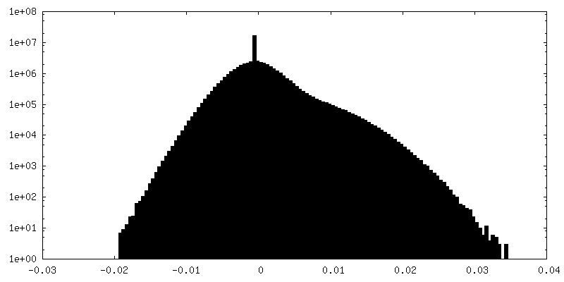
| Density Histograms |
- Sample components
-Entire : Cryo-EM structue of DIP-2I8I fibril polymorph 2
| Entire | Name: Cryo-EM structue of DIP-2I8I fibril polymorph 2 |
|---|---|
| Components |
|
-Supramolecule #1: Cryo-EM structue of DIP-2I8I fibril polymorph 2
| Supramolecule | Name: Cryo-EM structue of DIP-2I8I fibril polymorph 2 / type: complex / ID: 1 / Parent: 0 / Macromolecule list: all |
|---|---|
| Source (natural) | Organism: Homo sapiens (human) |
-Macromolecule #1: GLY-PHI-GLY-ASN-GLY-ASN-GLY-PHI-GLY
| Macromolecule | Name: GLY-PHI-GLY-ASN-GLY-ASN-GLY-PHI-GLY / type: protein_or_peptide / ID: 1 / Number of copies: 22 / Enantiomer: LEVO |
|---|---|
| Source (natural) | Organism: Homo sapiens (human) |
| Molecular weight | Theoretical: 1.077621 KDa |
| Sequence | String: G(PHI)GNGNG(PHI)G |
-Experimental details
-Structure determination
| Method | cryo EM |
|---|---|
 Processing Processing | helical reconstruction |
| Aggregation state | filament |
-Sample preparation
| Buffer | pH: 7 |
|---|---|
| Vitrification | Cryogen name: ETHANE |
- Electron microscopy
| Microscope | FEI TITAN KRIOS |
|---|---|
| Electron beam | Acceleration voltage: 300 kV / Electron source: FIELD EMISSION GUN |
| Electron optics | Illumination mode: FLOOD BEAM / Imaging mode: BRIGHT FIELDBright-field microscopy / Nominal defocus max: 2.0 µm / Nominal defocus min: 1.0 µm |
| Image recording | Film or detector model: GATAN K3 BIOQUANTUM (6k x 4k) / Average electron dose: 60.0 e/Å2 |
| Experimental equipment |  Model: Titan Krios / Image courtesy: FEI Company |
-Image processing
| Startup model | Type of model: NONE |
|---|---|
| Final angle assignment | Type: NOT APPLICABLE |
| Final reconstruction | Applied symmetry - Helical parameters - Δz: 4.83 Å Applied symmetry - Helical parameters - Δ&Phi: -1.21 ° Applied symmetry - Helical parameters - Axial symmetry: C1 (asymmetric) Resolution.type: BY AUTHOR / Resolution: 3.75 Å / Resolution method: FSC 0.143 CUT-OFF / Number images used: 11823 |
| FSC plot (resolution estimation) | 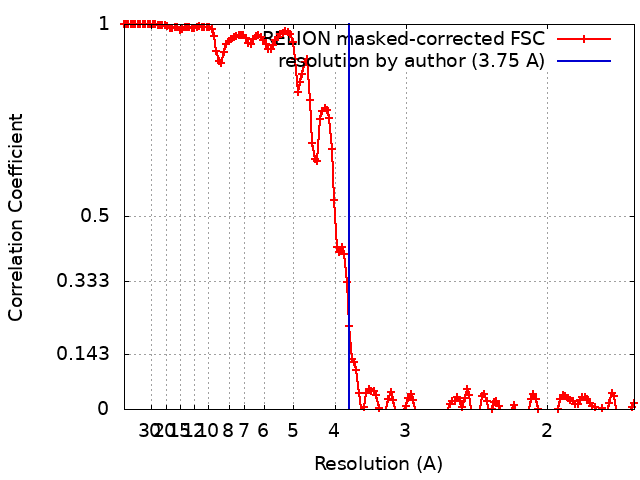 |
