 Movie
Movie Controller

[English] 日本語
 Yorodumi
- EMDB-35600: Cryo-Em structure of the middle part of the shrimp white spot syn... -
+ Open data

- Basic information
| Entry | 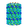 | |||||||||
|---|---|---|---|---|---|---|---|---|---|---|
| Title | Cryo-Em structure of the middle part of the shrimp white spot syndrome virus nucleocapsid (narrow type) | |||||||||
 Map data Map data | ||||||||||
 Sample Sample |
| |||||||||
| Biological species |   White spot syndrome virus White spot syndrome virus | |||||||||
| Method | single particle reconstruction / cryo EM / Resolution: 9.5 Å | |||||||||
 Authors Authors | Huang HJ / Wang HC | |||||||||
| Funding support |  Taiwan, 2 items Taiwan, 2 items
| |||||||||
 Citation Citation | Journal: Int J Mol Sci / Year: 2023 Title: Multiple Nucleocapsid Structural Forms of Shrimp White Spot Syndrome Virus Suggests a Novel Viral Morphogenetic Pathway. Authors: Hui-Ju Huang / Sen-Lin Tang / Yuan-Chih Chang / Hao-Ching Wang / Tze Hann Ng / Rees F Garmann / Yu-Wen Chen / Jiun-Yan Huang / Ramya Kumar / Sheng-Hsiung Chang / Shang-Rung Wu / Chih-Yu Chao ...Authors: Hui-Ju Huang / Sen-Lin Tang / Yuan-Chih Chang / Hao-Ching Wang / Tze Hann Ng / Rees F Garmann / Yu-Wen Chen / Jiun-Yan Huang / Ramya Kumar / Sheng-Hsiung Chang / Shang-Rung Wu / Chih-Yu Chao / Kyoko Matoba / Iwasaki Kenji / William M Gelbart / Tzu-Ping Ko / Hwei-Jiung Andrew Wang / Chu-Fang Lo / Li-Li Chen / Han-Ching Wang /    Abstract: White spot syndrome virus (WSSV) is a very large dsDNA virus. The accepted shape of the WSSV virion has been as ellipsoidal, with a tail-like extension. However, due to the scarcity of reliable ...White spot syndrome virus (WSSV) is a very large dsDNA virus. The accepted shape of the WSSV virion has been as ellipsoidal, with a tail-like extension. However, due to the scarcity of reliable references, the pathogenesis and morphogenesis of WSSV are not well understood. Here, we used transmission electron microscopy (TEM) and cryogenic electron microscopy (Cryo-EM) to address some knowledge gaps. We concluded that mature WSSV virions with a stout oval-like shape do not have tail-like extensions. Furthermore, there were two distinct ends in WSSV nucleocapsids: a portal cap and a closed base. A C14 symmetric structure of the WSSV nucleocapsid was also proposed, according to our Cryo-EM map. Immunoelectron microscopy (IEM) revealed that VP664 proteins, the main components of the 14 assembly units, form a ring-like architecture. Moreover, WSSV nucleocapsids were also observed to undergo unique helical dissociation. Based on these new results, we propose a novel morphogenetic pathway of WSSV. | |||||||||
| History |
|
- Structure visualization
| Supplemental images |
|---|

- Downloads & links
-EMDB archive
| Map data | emd_35600.map.gz | 195.9 MB |  EMDB map data format EMDB map data format | |
|---|---|---|---|---|
| Header (meta data) | emd-35600-v30.xmlemd-35600.xml | 15.8 KB 15.8 KB | Display Display | EMDB header |
| Images | 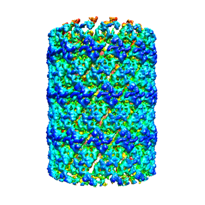 emd_35600.png emd_35600.png | 164.1 KB | ||
| Others | emd_35600_additional_1.map.gzemd_35600_half_map_1.map.gzemd_35600_half_map_2.map.gz | 1.4 GB 1.4 GB 1.4 GB | ||
| Archive directory |  http://ftp.pdbj.org/pub/emdb/structures/EMD-35600ftp://ftp.pdbj.org/pub/emdb/structures/EMD-35600 http://ftp.pdbj.org/pub/emdb/structures/EMD-35600ftp://ftp.pdbj.org/pub/emdb/structures/EMD-35600 | HTTPS FTP |
-Related structure data
-Links
| EMDB pages | EMDB (EBI/PDBe) / EMDataResource |
|---|
-Map
| File | Download / File: emd_35600.map.gz / Format: CCP4 / Size: 1.7 GB / Type: IMAGE STORED AS FLOATING POINT NUMBER (4 BYTES) | ||||||||||||||||||||
|---|---|---|---|---|---|---|---|---|---|---|---|---|---|---|---|---|---|---|---|---|---|
| Voxel size | X=Y=Z: 1.38 Å | ||||||||||||||||||||
| Density |
| ||||||||||||||||||||
| Symmetry | Space group: 1 | ||||||||||||||||||||
| Details | EMDB XML:
|
-Supplemental data
-Additional map: #1
| File | emd_35600_additional_1.map | ||||||||||||
|---|---|---|---|---|---|---|---|---|---|---|---|---|---|
| Projections & Slices |
| ||||||||||||
| Density Histograms |
 Z
Z Y
Y X
X


-Half map: #1
| File | emd_35600_half_map_1.map | ||||||||||||
|---|---|---|---|---|---|---|---|---|---|---|---|---|---|
| Projections & Slices |
| ||||||||||||
| Density Histograms |


-Half map: #2
| File | emd_35600_half_map_2.map | ||||||||||||
|---|---|---|---|---|---|---|---|---|---|---|---|---|---|
| Projections & Slices |
| ||||||||||||
| Density Histograms |


- Sample components
-Entire : White spot syndrome virus
| Entire | Name: White spot syndrome virus |
|---|---|
| Components |
|
-Supramolecule #1: White spot syndrome virus
| Supramolecule | Name: White spot syndrome virus / type: virus / ID: 1 / Parent: 0 / NCBI-ID: 342409 / Sci species name: White spot syndrome virus / Virus type: VIRION / Virus isolate: STRAIN / Virus enveloped: No / Virus empty: Yes |
|---|---|
| Host (natural) | Organism: Shrimp white spot syndrome virus / Strain: AF440570 |
-Experimental details
-Structure determination
| Method | cryo EM |
|---|---|
 Processing Processing | single particle reconstruction |
| Aggregation state | particle |
-Sample preparation
| Buffer | pH: 7.5 |
|---|---|
| Grid | Model: Quantifoil R1.2/1.3 / Material: COPPER / Mesh: 200 / Pretreatment - Type: GLOW DISCHARGE / Pretreatment - Time: 15 sec. |
| Vitrification | Cryogen name: ETHANE / Chamber humidity: 100 % / Chamber temperature: 274 K |
- Electron microscopy
| Microscope | FEI TALOS ARCTICA |
|---|---|
| Electron beam | Acceleration voltage: 200 kV / Electron source: FIELD EMISSION GUN |
| Electron optics | C2 aperture diameter: 50.0 µm / Calibrated magnification: 73000 / Illumination mode: FLOOD BEAM / Imaging mode: BRIGHT FIELDBright-field microscopy / Nominal defocus max: 2.5 µm / Nominal defocus min: 0.5 µm |
| Sample stage | Specimen holder model: OTHER / Cooling holder cryogen: NITROGEN |
| Image recording | Film or detector model: FEI FALCON III (4k x 4k) / Average electron dose: 48.0 e/Å2 |
| Experimental equipment |  Model: Talos Arctica / Image courtesy: FEI Company |
-Image processing
| Initial angle assignment | Type: PROJECTION MATCHING |
|---|---|
| Final angle assignment | Type: PROJECTION MATCHING |
| Final reconstruction | Resolution.type: BY AUTHOR / Resolution: 9.5 Å / Resolution method: FSC 0.143 CUT-OFF / Number images used: 15279 |
-Atomic model buiding 1
| Refinement | Protocol: AB INITIO MODEL |
|---|
