 Movie
Movie Controller

[English] 日本語
 Yorodumi
- EMDB-3554: Yeast mitochondrial ribosome - small subunit head (fitted to EMD 3551) -
+ Open data

- Basic information
| Entry | Database: EMDB / ID: EMD-3554 | |||||||||
|---|---|---|---|---|---|---|---|---|---|---|
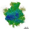
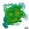
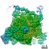
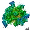
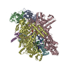
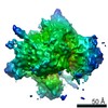
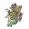
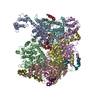
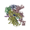
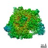
| Title | Yeast mitochondrial ribosome - small subunit head (fitted to EMD 3551) | |||||||||
 Map data Map data | ||||||||||
 Sample Sample |
| |||||||||
| Biological species |   Saccharomyces cerevisiae (brewer's yeast) Saccharomyces cerevisiae (brewer's yeast) | |||||||||
| Method | single particle reconstruction / cryo EM / Resolution: 3.5 Å | |||||||||
 Authors Authors | Desai N / Brown A / Amunts A / Ramakrishnan V | |||||||||
 Citation Citation | Journal: Science / Year: 2017 Title: The structure of the yeast mitochondrial ribosome. Authors: Nirupa Desai / Alan Brown / Alexey Amunts / V Ramakrishnan /   Abstract: Mitochondria have specialized ribosomes (mitoribosomes) dedicated to the expression of the genetic information encoded by their genomes. Here, using electron cryomicroscopy, we have determined the ...Mitochondria have specialized ribosomes (mitoribosomes) dedicated to the expression of the genetic information encoded by their genomes. Here, using electron cryomicroscopy, we have determined the structure of the 75-component yeast mitoribosome to an overall resolution of 3.3 angstroms. The mitoribosomal small subunit has been built de novo and includes 15S ribosomal RNA (rRNA) and 34 proteins, including 14 without homologs in the evolutionarily related bacterial ribosome. Yeast-specific rRNA and protein elements, including the acquisition of a putatively active enzyme, give the mitoribosome a distinct architecture compared to the mammalian mitoribosome. At an expanded messenger RNA channel exit, there is a binding platform for translational activators that regulate translation in yeast but not mammalian mitochondria. The structure provides insights into the evolution and species-specific specialization of mitochondrial translation. | |||||||||
| History |
|
- Structure visualization
| Movie |
 Movie viewer Movie viewer |
|---|---|
| Structure viewer | EM map: SurfViewMolmilJmol/JSmol |
| Supplemental images |

- Downloads & links
-EMDB archive
| Map data | emd_3554.map.gz | 2.4 MB | EMDB map data format | |
|---|---|---|---|---|
| Header (meta data) | emd-3554-v30.xmlemd-3554.xml | 12.1 KB 12.1 KB | Display Display | EMDB header |
| Images | 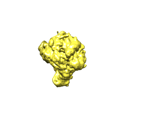 emd_3554.png emd_3554.png | 52.4 KB | ||
| Others | emd_3554_half_map_1.map.gzemd_3554_half_map_2.map.gz | 117.9 MB 117.1 MB | ||
| Archive directory |  http://ftp.pdbj.org/pub/emdb/structures/EMD-3554ftp://ftp.pdbj.org/pub/emdb/structures/EMD-3554 http://ftp.pdbj.org/pub/emdb/structures/EMD-3554ftp://ftp.pdbj.org/pub/emdb/structures/EMD-3554 | HTTPS FTP |
-Related structure data
| Related structure data |  3551C  3552C  3553C  3555C  3556C  5mrcC 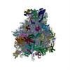 5mreC 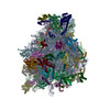 5mrfC C: citing same article ( |
|---|---|
| Similar structure data | |
| EM raw data | EMPIAR-10107 (Title: The Structure of the Yeast Mitochondrial Ribosome / Data size: 138.6 Data #1: Yeast mitochondrial ribosome - shiny particles [picked particles - multiframe - processed]) |

-Links
| EMDB pages | EMDB (EBI/PDBe) / EMDataResource |
|---|---|
| Related items in Molecule of the Month |

-Map
| File | Download / File: emd_3554.map.gz / Format: CCP4 / Size: 137.1 MB / Type: IMAGE STORED AS FLOATING POINT NUMBER (4 BYTES) | ||||||||||||||||||||||||||||||||||||||||||||||||||||||||||||
|---|---|---|---|---|---|---|---|---|---|---|---|---|---|---|---|---|---|---|---|---|---|---|---|---|---|---|---|---|---|---|---|---|---|---|---|---|---|---|---|---|---|---|---|---|---|---|---|---|---|---|---|---|---|---|---|---|---|---|---|---|---|
| Voxel size | X=Y=Z: 1.34 Å | ||||||||||||||||||||||||||||||||||||||||||||||||||||||||||||
| Density |
| ||||||||||||||||||||||||||||||||||||||||||||||||||||||||||||
| Symmetry | Space group: 1 | ||||||||||||||||||||||||||||||||||||||||||||||||||||||||||||
| Details | EMDB XML:
CCP4 map header:
| ||||||||||||||||||||||||||||||||||||||||||||||||||||||||||||
-Supplemental data
-Half map: #1
| File | emd_3554_half_map_1.map | ||||||||||||
|---|---|---|---|---|---|---|---|---|---|---|---|---|---|
| Projections & Slices |
| ||||||||||||
| Density Histograms |
 Z
Z Y
Y X
X


-Half map: #2
| File | emd_3554_half_map_2.map | ||||||||||||
|---|---|---|---|---|---|---|---|---|---|---|---|---|---|
| Projections & Slices |
| ||||||||||||
| Density Histograms |


- Sample components
-Entire : Yeast mitochondrial ribosome - small subunit head
| Entire | Name: Yeast mitochondrial ribosome - small subunit head |
|---|---|
| Components |
|
-Supramolecule #1: Yeast mitochondrial ribosome - small subunit head
| Supramolecule | Name: Yeast mitochondrial ribosome - small subunit head / type: complex / ID: 1 / Parent: 0 |
|---|---|
| Source (natural) | Organism: Saccharomyces cerevisiae (brewer's yeast) |
-Experimental details
-Structure determination
| Method | cryo EM |
|---|---|
 Processing Processing | single particle reconstruction |
| Aggregation state | particle |
-Sample preparation
| Buffer | pH: 7.5 |
|---|---|
| Vitrification | Cryogen name: ETHANE |
- Electron microscopy
| Microscope | FEI TITAN KRIOS |
|---|---|
| Electron beam | Acceleration voltage: 300 kV / Electron source: FIELD EMISSION GUN |
| Electron optics | Illumination mode: FLOOD BEAM / Imaging mode: BRIGHT FIELDBright-field microscopy |
| Image recording | Film or detector model: FEI FALCON II (4k x 4k) / Average electron dose: 23.5 e/Å2 |
| Experimental equipment |  Model: Titan Krios / Image courtesy: FEI Company |
-Image processing
| CTF correction | Software - Name: RELION (ver. 1.4) |
|---|---|
| Initial angle assignment | Type: OTHER / Software - Name: RELION (ver. 1.4) |
| Final angle assignment | Type: ANGULAR RECONSTITUTION / Software - Name: RELION (ver. 1.4) |
| Final reconstruction | Resolution.type: BY AUTHOR / Resolution: 3.5 Å / Resolution method: FSC 0.143 CUT-OFF / Software - Name: RELION (ver. 1.4) / Number images used: 264961 |
