 Movie
Movie Controller

[English] 日本語
 Yorodumi
- EMDB-3547: Cryo-EM structure of a human spliceosome activated for step 2 of ... -
+ Open data

- Basic information
| Entry | Database: EMDB / ID: EMD-3547 | |||||||||
|---|---|---|---|---|---|---|---|---|---|---|
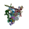
| Title | Cryo-EM structure of a human spliceosome activated for step 2 of splicing (C* complex) | |||||||||
 Map data Map data | ||||||||||
 Sample Sample |
| |||||||||
| Biological species |   Homo sapiens (human) Homo sapiens (human) | |||||||||
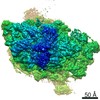
| Method | single particle reconstruction / cryo EM / Resolution: 8.4 Å | |||||||||
 Authors Authors | Bertram K / Liu WT | |||||||||
 Citation Citation | Journal: Nature / Year: 2017 Title: Cryo-EM structure of a human spliceosome activated for step 2 of splicing. Authors: Karl Bertram / Dmitry E Agafonov / Wen-Ti Liu / Olexandr Dybkov / Cindy L Will / Klaus Hartmuth / Henning Urlaub / Berthold Kastner / Holger Stark / Reinhard Lührmann /  Abstract: Spliceosome rearrangements facilitated by RNA helicase PRP16 before catalytic step two of splicing are poorly understood. Here we report a 3D cryo-electron microscopy structure of the human ...Spliceosome rearrangements facilitated by RNA helicase PRP16 before catalytic step two of splicing are poorly understood. Here we report a 3D cryo-electron microscopy structure of the human spliceosomal C complex stalled directly after PRP16 action (C*). The architecture of the catalytic U2-U6 ribonucleoprotein (RNP) core of the human C* spliceosome is very similar to that of the yeast pre-Prp16 C complex. However, in C* the branched intron region is separated from the catalytic centre by approximately 20 Å, and its position close to the U6 small nuclear RNA ACAGA box is stabilized by interactions with the PRP8 RNase H-like and PRP17 WD40 domains. RNA helicase PRP22 is located about 100 Å from the catalytic centre, suggesting that it destabilizes the spliced mRNA after step two from a distance. Comparison of the structure of the yeast C and human C* complexes reveals numerous RNP rearrangements that are likely to be facilitated by PRP16, including a large-scale movement of the U2 small nuclear RNP. | |||||||||
| History |
|
- Structure visualization
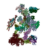
| Movie |
 Movie viewer Movie viewer |
|---|---|
| Structure viewer | EM map: SurfViewMolmilJmol/JSmol |
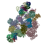
| Supplemental images |

- Downloads & links
-EMDB archive
| Map data | emd_3547.map.gz | 162.1 MB | EMDB map data format | |
|---|---|---|---|---|
| Header (meta data) | emd-3547-v30.xmlemd-3547.xml | 18.1 KB 18.1 KB | Display Display | EMDB header |
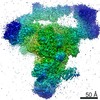
| Images | 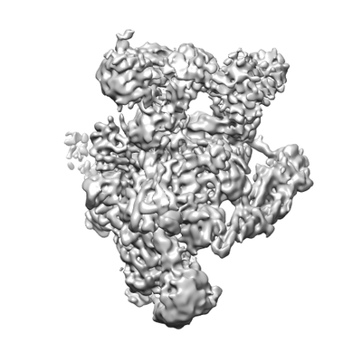 emd_3547.png emd_3547.png | 53.5 KB | ||
| Others | emd_3547_half_map_1.map.gzemd_3547_half_map_2.map.gz | 139.5 MB 139.5 MB | ||
| Archive directory |  http://ftp.pdbj.org/pub/emdb/structures/EMD-3547ftp://ftp.pdbj.org/pub/emdb/structures/EMD-3547 http://ftp.pdbj.org/pub/emdb/structures/EMD-3547ftp://ftp.pdbj.org/pub/emdb/structures/EMD-3547 | HTTPS FTP |
-Related structure data


-Links
| EMDB pages | EMDB (EBI/PDBe) / EMDataResource |
|---|
-Map
| File | Download / File: emd_3547.map.gz / Format: CCP4 / Size: 178 MB / Type: IMAGE STORED AS FLOATING POINT NUMBER (4 BYTES) | ||||||||||||||||||||||||||||||||||||||||||||||||||||||||||||
|---|---|---|---|---|---|---|---|---|---|---|---|---|---|---|---|---|---|---|---|---|---|---|---|---|---|---|---|---|---|---|---|---|---|---|---|---|---|---|---|---|---|---|---|---|---|---|---|---|---|---|---|---|---|---|---|---|---|---|---|---|---|
| Voxel size | X=Y=Z: 1.59 Å | ||||||||||||||||||||||||||||||||||||||||||||||||||||||||||||
| Density |
| ||||||||||||||||||||||||||||||||||||||||||||||||||||||||||||
| Symmetry | Space group: 1 | ||||||||||||||||||||||||||||||||||||||||||||||||||||||||||||
| Details | EMDB XML:
CCP4 map header:
| ||||||||||||||||||||||||||||||||||||||||||||||||||||||||||||
-Supplemental data
-Half map: #1
| File | emd_3547_half_map_1.map | ||||||||||||
|---|---|---|---|---|---|---|---|---|---|---|---|---|---|
| Projections & Slices |
| ||||||||||||
| Density Histograms |
 Z
Z Y
Y X
X


-Half map: #2
| File | emd_3547_half_map_2.map | ||||||||||||
|---|---|---|---|---|---|---|---|---|---|---|---|---|---|
| Projections & Slices |
| ||||||||||||
| Density Histograms |


- Sample components
-Entire : Human C* Spliceosome, unmasked refinement
| Entire | Name: Human C* Spliceosome, unmasked refinement |
|---|---|
| Components |
|
-Supramolecule #1: Human C* Spliceosome, unmasked refinement
| Supramolecule | Name: Human C* Spliceosome, unmasked refinement / type: complex / ID: 1 / Parent: 0 / Macromolecule list: #1-#41 |
|---|---|
| Source (natural) | Organism: Homo sapiens (human) |
-Experimental details
-Structure determination
| Method | cryo EM |
|---|---|
 Processing Processing | single particle reconstruction |
| Aggregation state | particle |
-Sample preparation
| Buffer | pH: 6.4 |
|---|---|
| Vitrification | Cryogen name: ETHANE / Chamber humidity: 80 % / Chamber temperature: 277 K |
- Electron microscopy
| Microscope | FEI TITAN KRIOS |
|---|---|
| Electron beam | Acceleration voltage: 300 kV / Electron source: FIELD EMISSION GUN |
| Electron optics | Illumination mode: SPOT SCAN / Imaging mode: BRIGHT FIELDBright-field microscopy / Cs: 0.01 mm |
| Image recording | Film or detector model: FEI FALCON II (4k x 4k) / Digitization - Frames/image: 2-17 / Average electron dose: 2.1 e/Å2 |
| Experimental equipment |  Model: Titan Krios / Image courtesy: FEI Company |
-Image processing
| Startup model | Type of model: OTHER / Details: Reconstruction from negatively stained particles |
|---|---|
| Initial angle assignment | Type: COMMON LINE |
| Final angle assignment | Type: OTHER |
| Final reconstruction | Applied symmetry - Point group: C1 (asymmetric) / Resolution.type: BY AUTHOR / Resolution: 8.4 Å / Resolution method: FSC 0.143 CUT-OFF / Number images used: 136534 |
-Atomic model buiding 1
| Refinement | Space: REAL |
|---|
