 Movie
Movie Controller


+ Open data

- Basic information
| Entry | Database: EMDB / ID: EMD-3509 | |||||||||
|---|---|---|---|---|---|---|---|---|---|---|
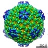
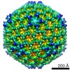
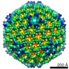
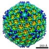
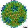
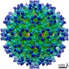
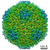
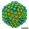
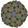
| Title | IBDV E1-1B population | |||||||||
 Map data Map data | ||||||||||
 Sample Sample |
| |||||||||
| Biological species |  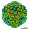  Infectious bursal disease virus (Gumboro virus) Infectious bursal disease virus (Gumboro virus) | |||||||||
| Method | single particle reconstruction / cryo EM / Resolution: 20.6 Å | |||||||||
 Authors Authors | Mata CP / Mertens J / Fontana J / Luque D / Allende-Ballestero C / Reguera D / Trus BL / Steven AC / Carrascosa JL / Caston JR | |||||||||
 Citation Citation | Journal: J Virol / Year: 2018 Title: The RNA-Binding Protein of a Double-Stranded RNA Virus Acts like a Scaffold Protein. Authors: Carlos P Mata / Johann Mertens / Juan Fontana / Daniel Luque / Carolina Allende-Ballestero / David Reguera / Benes L Trus / Alasdair C Steven / José L Carrascosa / José R Castón /   Abstract: Infectious bursal disease virus (IBDV), a nonenveloped, double-stranded RNA (dsRNA) virus with a T=13 icosahedral capsid, has a virion assembly strategy that initiates with a precursor particle based ...Infectious bursal disease virus (IBDV), a nonenveloped, double-stranded RNA (dsRNA) virus with a T=13 icosahedral capsid, has a virion assembly strategy that initiates with a precursor particle based on an internal scaffold shell similar to that of tailed double-stranded DNA (dsDNA) viruses. In IBDV-infected cells, the assembly pathway results mainly in mature virions that package four dsRNA segments, although minor viral populations ranging from zero to three dsRNA segments also form. We used cryo-electron microscopy (cryo-EM), cryo-electron tomography, and atomic force microscopy to characterize these IBDV populations. The VP3 protein was found to act as a scaffold protein by building an irregular, ∼40-Å-thick internal shell without icosahedral symmetry, which facilitates formation of a precursor particle, the procapsid. Analysis of IBDV procapsid mechanical properties indicated a VP3 layer beneath the icosahedral shell, which increased the effective capsid thickness. Whereas scaffolding proteins are discharged in tailed dsDNA viruses, VP3 is a multifunctional protein. In mature virions, VP3 is bound to the dsRNA genome, which is organized as ribonucleoprotein complexes. IBDV is an amalgam of dsRNA viral ancestors and traits from dsDNA and single-stranded RNA (ssRNA) viruses. Structural analyses highlight the constraint of virus evolution to a limited number of capsid protein folds and assembly strategies that result in a functional virion. We report the cryo-EM and cryo-electron tomography structures and the results of atomic force microscopy studies of the infectious bursal disease virus (IBDV), a double-stranded RNA virus with an icosahedral capsid. We found evidence of a new inner shell that might act as an internal scaffold during IBDV assembly. The use of an internal scaffold is reminiscent of tailed dsDNA viruses, which constitute the most successful self-replicating system on Earth. The IBDV scaffold protein is multifunctional and, after capsid maturation, is genome bound to form ribonucleoprotein complexes. IBDV encompasses numerous functional and structural characteristics of RNA and DNA viruses; we suggest that IBDV is a modern descendant of ancestral viruses and comprises different features of current viral lineages. | |||||||||
| History |
|
- Structure visualization
| Movie |
 Movie viewer Movie viewer |
|---|---|
| Structure viewer | EM map: SurfViewMolmilJmol/JSmol |
| Supplemental images |

- Downloads & links
-EMDB archive
| Map data | emd_3509.map.gz | 15.3 MB | EMDB map data format | |
|---|---|---|---|---|
| Header (meta data) | emd-3509-v30.xmlemd-3509.xml | 11.2 KB 11.2 KB | Display Display | EMDB header |
| Images | 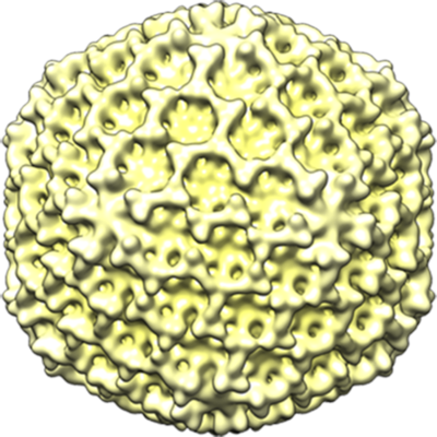 emd_3509.png emd_3509.png | 252.5 KB | ||
| Archive directory |  http://ftp.pdbj.org/pub/emdb/structures/EMD-3509ftp://ftp.pdbj.org/pub/emdb/structures/EMD-3509 http://ftp.pdbj.org/pub/emdb/structures/EMD-3509ftp://ftp.pdbj.org/pub/emdb/structures/EMD-3509 | HTTPS FTP |
-Related structure data
| Related structure data | 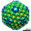 3507C 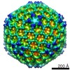 3510C 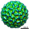 3511C 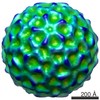 3512C 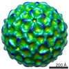 3513C 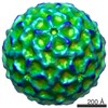 3514C 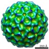 3515C  3516C C: citing same article ( |
|---|---|
| Similar structure data |
-Links
| EMDB pages | EMDB (EBI/PDBe) / EMDataResource |
|---|
-Map
| File | Download / File: emd_3509.map.gz / Format: CCP4 / Size: 30.5 MB / Type: IMAGE STORED AS FLOATING POINT NUMBER (4 BYTES) | ||||||||||||||||||||||||||||||||||||||||||||||||||||||||||||
|---|---|---|---|---|---|---|---|---|---|---|---|---|---|---|---|---|---|---|---|---|---|---|---|---|---|---|---|---|---|---|---|---|---|---|---|---|---|---|---|---|---|---|---|---|---|---|---|---|---|---|---|---|---|---|---|---|---|---|---|---|---|
| Voxel size | X=Y=Z: 4.32 Å | ||||||||||||||||||||||||||||||||||||||||||||||||||||||||||||
| Density |
| ||||||||||||||||||||||||||||||||||||||||||||||||||||||||||||
| Symmetry | Space group: 1 | ||||||||||||||||||||||||||||||||||||||||||||||||||||||||||||
| Details | EMDB XML:
CCP4 map header:
| ||||||||||||||||||||||||||||||||||||||||||||||||||||||||||||
-Supplemental data
- Sample components
-Entire : Infectious bursal disease virus
| Entire | Name: Infectious bursal disease virus (Gumboro virus) |
|---|---|
| Components |
|
-Supramolecule #1: Infectious bursal disease virus
| Supramolecule | Name: Infectious bursal disease virus / type: virus / ID: 1 / Parent: 0 / NCBI-ID: 10995 / Sci species name: Infectious bursal disease virus / Virus type: VIRION / Virus isolate: STRAIN / Virus enveloped: No / Virus empty: Yes |
|---|---|
| Host (natural) | Organism:  Gallus gallus (chicken) Gallus gallus (chicken) |
| Molecular weight | Experimental: 50 MDa |
| Virus shell | Shell ID: 1 / Name: VP2 / Diameter: 700.0 Å / T number (triangulation number): 13 |
-Experimental details
-Structure determination
| Method | cryo EM |
|---|---|
 Processing Processing | single particle reconstruction |
| Aggregation state | particle |
-Sample preparation
| Buffer | pH: 6.2 / Details: 25 mM PIPES pH 6.2, 150 mM NaCl, 20 mM CaCl2 |
|---|---|
| Grid | Model: Quantifoil R2/2 / Material: COPPER/RHODIUM / Mesh: 300 |
| Vitrification | Cryogen name: ETHANE / Instrument: LEICA EM CPC |
- Electron microscopy
| Microscope | FEI TECNAI F20 |
|---|---|
| Electron beam | Acceleration voltage: 200 kV / Electron source: FIELD EMISSION GUN |
| Electron optics | Calibrated defocus max: 5.0 µm / Calibrated defocus min: 0.7 µm / Illumination mode: FLOOD BEAM / Imaging mode: BRIGHT FIELDBright-field microscopy / Cs: 2.26 mm / Nominal magnification: 50000 |
| Sample stage | Specimen holder model: GATAN 626 SINGLE TILT LIQUID NITROGEN CRYO TRANSFER HOLDER Cooling holder cryogen: NITROGEN |
| Image recording | Film or detector model: FEI EAGLE (4k x 4k) / Average electron dose: 10.0 e/Å2 |
| Experimental equipment |  Model: Tecnai F20 / Image courtesy: FEI Company |
-Image processing
| Particle selection | Number selected: 5565 Details: Includes E1-1A and E1-1B particles further classified |
|---|---|
| CTF correction | Software - Name: CTFFIND (ver. 3) |
| Startup model | Type of model: EMDB MAP EMDB ID: |
| Initial angle assignment | Type: PROJECTION MATCHING / Software - Name: RELION (ver. 1.4) |
| Final 3D classification | Number classes: 2 / Avg.num./class: 2027 / Software - Name: RELION (ver. 1.4) |
| Final angle assignment | Type: PROJECTION MATCHING / Software - Name: RELION (ver. 1.4) |
| Final reconstruction | Number classes used: 1 / Applied symmetry - Point group: I (icosahedral) / Algorithm: FOURIER SPACE / Resolution.type: BY AUTHOR / Resolution: 20.6 Å / Resolution method: FSC 0.33 CUT-OFF / Software - Name: RELION (ver. 1.4) / Number images used: 2027 |
