 Movie
Movie Controller

[English] 日本語
 Yorodumi
- EMDB-3455: Thermoplasma acidophilum 20S proteasome map reconstructed with Re... -
+ Open data

- Basic information
| Entry | Database: EMDB / ID: EMD-3455 | |||||||||
|---|---|---|---|---|---|---|---|---|---|---|
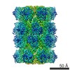
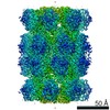
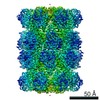
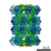
| Title | Thermoplasma acidophilum 20S proteasome map reconstructed with Relion 2.0 particle polishing from defocused Volta phase plate cryo-EM data | |||||||||
 Map data Map data | Thermoplasma acidophilum 20S proteasome map reconstructed with Relion 2.0 particle polishing from defocused Volta phase plate cryo-EM data. | |||||||||
 Sample Sample |
| |||||||||
| Biological species |    Thermoplasma acidophilum (acidophilic) Thermoplasma acidophilum (acidophilic) | |||||||||
| Method | single particle reconstruction / cryo EM / Resolution: 2.4 Å | |||||||||
 Authors Authors | Danev R / Tegunov D / Baumeister W | |||||||||
 Citation Citation | Journal: Elife / Year: 2017 Title: Using the Volta phase plate with defocus for cryo-EM single particle analysis. Authors: Radostin Danev / Dimitry Tegunov / Wolfgang Baumeister /  Abstract: Previously, we reported an in-focus data acquisition method for cryo-EM single-particle analysis with the Volta phase plate (Danev and Baumeister, 2016). Here, we extend the technique to include a ...Previously, we reported an in-focus data acquisition method for cryo-EM single-particle analysis with the Volta phase plate (Danev and Baumeister, 2016). Here, we extend the technique to include a small amount of defocus which enables contrast transfer function measurement and correction. This hybrid approach simplifies the experiment and increases the data acquisition speed. It also removes the resolution limit inherent to the in-focus method thus allowing 3D reconstructions with resolutions better than 3 Å. | |||||||||
| History |
|
- Structure visualization
| Movie |
 Movie viewer Movie viewer |
|---|---|
| Structure viewer | EM map: SurfViewMolmilJmol/JSmol |
| Supplemental images |

- Downloads & links
-EMDB archive
| Map data | emd_3455.map.gz | 165.5 MB | EMDB map data format | |
|---|---|---|---|---|
| Header (meta data) | emd-3455-v30.xmlemd-3455.xml | 12.6 KB 12.6 KB | Display Display | EMDB header |
| Images | 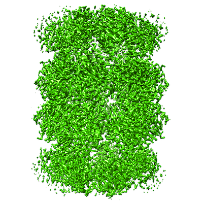 emd_3455.png emd_3455.png | 238.8 KB | ||
| Archive directory |  http://ftp.pdbj.org/pub/emdb/structures/EMD-3455ftp://ftp.pdbj.org/pub/emdb/structures/EMD-3455 http://ftp.pdbj.org/pub/emdb/structures/EMD-3455ftp://ftp.pdbj.org/pub/emdb/structures/EMD-3455 | HTTPS FTP |
-Related structure data
| Related structure data |  3456C  3457C C: citing same article ( |
|---|---|
| Similar structure data | |
| EM raw data | EMPIAR-10078 (Title: Volta phase plate with defocus cryo-EM dataset of Thermoplasma acidophilum 20S proteasome Data size: 92.5 Data #1: Unaligned multi-frame micrographs of 20S proteasome acquired with a Volta phase plate and defocus [micrographs - multiframe]) |

-Links
| EMDB pages | EMDB (EBI/PDBe) / EMDataResource |
|---|
-Map
| File | Download / File: emd_3455.map.gz / Format: CCP4 / Size: 178 MB / Type: IMAGE STORED AS FLOATING POINT NUMBER (4 BYTES) | ||||||||||||||||||||||||||||||||||||||||||||||||||||||||||||||||||||
|---|---|---|---|---|---|---|---|---|---|---|---|---|---|---|---|---|---|---|---|---|---|---|---|---|---|---|---|---|---|---|---|---|---|---|---|---|---|---|---|---|---|---|---|---|---|---|---|---|---|---|---|---|---|---|---|---|---|---|---|---|---|---|---|---|---|---|---|---|---|
| Annotation | Thermoplasma acidophilum 20S proteasome map reconstructed with Relion 2.0 particle polishing from defocused Volta phase plate cryo-EM data. | ||||||||||||||||||||||||||||||||||||||||||||||||||||||||||||||||||||
| Voxel size | X=Y=Z: 0.527 Å | ||||||||||||||||||||||||||||||||||||||||||||||||||||||||||||||||||||
| Density |
| ||||||||||||||||||||||||||||||||||||||||||||||||||||||||||||||||||||
| Symmetry | Space group: 1 | ||||||||||||||||||||||||||||||||||||||||||||||||||||||||||||||||||||
| Details | EMDB XML:
CCP4 map header:
| ||||||||||||||||||||||||||||||||||||||||||||||||||||||||||||||||||||
-Supplemental data
- Sample components
-Entire : Thermoplasma acidophilum 20S proteasome
| Entire | Name: Thermoplasma acidophilum 20S proteasome |
|---|---|
| Components |
|
-Supramolecule #1: Thermoplasma acidophilum 20S proteasome
| Supramolecule | Name: Thermoplasma acidophilum 20S proteasome / type: complex / ID: 1 / Parent: 0 |
|---|---|
| Source (natural) | Organism: Thermoplasma acidophilum (acidophilic) |
| Recombinant expression | Organism:  Escherichia coli 'BL21-Gold(DE3)pLysS AG' (bacteria) Escherichia coli 'BL21-Gold(DE3)pLysS AG' (bacteria)Recombinant plasmid: pT7-7 |
| Molecular weight | Theoretical: 700 KDa |
-Experimental details
-Structure determination
| Method | cryo EM |
|---|---|
 Processing Processing | single particle reconstruction |
| Aggregation state | particle |
-Sample preparation
| Concentration | 0.5 mg/mL |
|---|---|
| Buffer | pH: 7.6 / Details: 25 mM Tris-HCL |
| Grid | Model: Quantifoil R1.2/1.3 / Material: COPPER / Mesh: 200 / Pretreatment - Type: GLOW DISCHARGE / Pretreatment - Atmosphere: OTHER / Pretreatment - Pressure: 0.1 kPa |
| Vitrification | Cryogen name: ETHANE-PROPANE / Chamber humidity: 95 % / Chamber temperature: 293 K / Instrument: FEI VITROBOT MARK III Details: 3 ul of 0.5 mg/ml protein solution was applied on a grid in the Vitrobot chamber set to 95% RH at 20 degC then blotted for 5 s and plunged. |
- Electron microscopy
| Microscope | FEI TITAN KRIOS |
|---|---|
| Electron beam | Acceleration voltage: 300 kV / Electron source: FIELD EMISSION GUN |
| Electron optics | C2 aperture diameter: 50.0 µm / Calibrated defocus max: 0.7 µm / Calibrated defocus min: 0.3 µm / Calibrated magnification: 47000 / Illumination mode: FLOOD BEAM / Imaging mode: BRIGHT FIELDBright-field microscopy / Cs: 2.62 mm / Nominal defocus max: 0.5 µm / Nominal defocus min: 0.3 µm / Nominal magnification: 47000 |
| Specialist optics | Phase plate: VOLTA PHASE PLATE / Energy filter - Name: GIF Quantum / Energy filter - Lower energy threshold: 0 eV / Energy filter - Upper energy threshold: 20 eV |
| Sample stage | Specimen holder model: FEI TITAN KRIOS AUTOGRID HOLDER / Cooling holder cryogen: NITROGEN |
| Image recording | Film or detector model: GATAN K2 SUMMIT (4k x 4k) / Detector mode: SUPER-RESOLUTION / Digitization - Frames/image: 1-24 / Number grids imaged: 1 / Number real images: 468 / Average exposure time: 12.0 sec. / Average electron dose: 39.0 e/Å2 |
| Experimental equipment |  Model: Titan Krios / Image courtesy: FEI Company |
-Image processing
| Particle selection | Number selected: 145870 |
|---|---|
| CTF correction | Software - Name: MATLAB / Software - details: homemade MATLAB program / Details: Relion 2.0 CTF correction |
| Startup model | Type of model: EMDB MAP EMDB ID: Details: 60 A low-pass filtered initial reference |
| Initial angle assignment | Type: PROJECTION MATCHING / Software - Name: RELION (ver. 2.0) |
| Final 3D classification | Number classes: 5 / Software - Name: RELION (ver. 2.0) |
| Final angle assignment | Type: PROJECTION MATCHING / Software - Name: RELION (ver. 2.0) |
| Final reconstruction | Number classes used: 1 / Applied symmetry - Point group: D7 (2x7 fold dihedral) / Resolution.type: BY AUTHOR / Resolution: 2.4 Å / Resolution method: FSC 0.143 CUT-OFF / Software - Name: RELION (ver. 2.0) / Number images used: 93596 |
-Atomic model buiding 1
| Refinement | Space: REAL / Protocol: RIGID BODY FIT / Overall B value: 74 |
|---|
