Journal: MedComm (2020) / Year: 2022 Title: Cryo-EM structure of G-protein-coupled receptor GPR17 in complex with inhibitory G protein. Authors: Fang Ye / Thian-Sze Wong / Geng Chen / Zhiyi Zhang / Binghao Zhang / Shiyi Gan / Wei Gao / Jiancheng Li / Zhangsong Wu / Xin Pan / Yang Du / Abstract: GPR17 is a class A orphan G protein-coupled receptor (GPCR) expressed in neurons and oligodendrocyte progenitors of the central nervous system (CNS). The signalling of GPR17 occurs through the ...GPR17 is a class A orphan G protein-coupled receptor (GPCR) expressed in neurons and oligodendrocyte progenitors of the central nervous system (CNS). The signalling of GPR17 occurs through the heterotrimeric Gi, but its activation mechanism is unclear. Here, we employed cryo-electron microscopy (cryo-EM) technology to elucidate the structure of activated GPR17-Gi complex. The 3.02 Å resolution structure, together with mutagenesis studies, revealed that the extracellular loop2 of GPR17 occupied the orthosteric binding pocket to promote its self-activation. The active GPR17 carried several typical microswitches like other class A GPCRs. Moreover, the Gi interacted with the key residues of transmembrane helix 3 (TM3), the amphipathic helix 8 (Helix8), and intracellular loops 3 (ICL3) in GPR17 to engage in the receptor core. In summary, our results highlight the activation mechanism of GPR17 from the structural basis. Elucidating the structural and activation mechanism of GPR17 may facilitate the pharmacological intervention for acute/chronic CNS injury.
In the structure databanks used in Yorodumi, some data are registered as the other names, "COVID-19 virus" and "2019-nCoV". Here are the details of the virus and the list of structure data.
Jan 31, 2019. EMDB accession codes are about to change! (news from PDBe EMDB page)
EMDB accession codes are about to change! (news from PDBe EMDB page)
The allocation of 4 digits for EMDB accession codes will soon come to an end. Whilst these codes will remain in use, new EMDB accession codes will include an additional digit and will expand incrementally as the available range of codes is exhausted. The current 4-digit format prefixed with “EMD-” (i.e. EMD-XXXX) will advance to a 5-digit format (i.e. EMD-XXXXX), and so on. It is currently estimated that the 4-digit codes will be depleted around Spring 2019, at which point the 5-digit format will come into force.
The EM Navigator/Yorodumi systems omit the EMD- prefix.
Related info.:Q: What is EMD? / ID/Accession-code notation in Yorodumi/EM Navigator
Yorodumi is a browser for structure data from EMDB, PDB, SASBDB, etc.
This page is also the successor to EM Navigator detail page, and also detail information page/front-end page for Omokage search.
The word "yorodu" (or yorozu) is an old Japanese word meaning "ten thousand". "mi" (miru) is to see.
Related info.:EMDB / PDB / SASBDB / Comparison of 3 databanks / Yorodumi Search / Aug 31, 2016. New EM Navigator & Yorodumi / Yorodumi Papers / Jmol/JSmol / Function and homology information / Changes in new EM Navigator and Yorodumi
 Movie
Movie Controller


 Open data
 Basic information
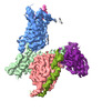
 Map data
 Sample
 Function and homology information
 P2Y receptors /

 Authors
 Citation
 Structure visualization
 Downloads & links
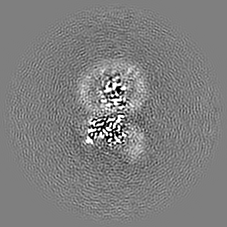
 emd_33682.png
 http://ftp.pdbj.org/pub/emdb/structures/EMD-33682
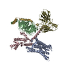


 Z
Z Y
Y X
X


 Sample components
 Processing
 Electron microscopy
