Movie
Movie Controller

[English] 日本語
 Yorodumi
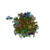
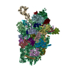
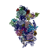
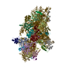
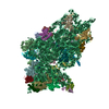
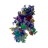
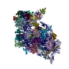
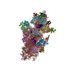
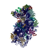
- EMDB-3300: Electron cryo-microscopy of a human pre-ribosomal (pre-40S) particle -
+ Open data

- Basic information
| Entry | Database: EMDB / ID: EMD-3300 | |||||||||
|---|---|---|---|---|---|---|---|---|---|---|
| Title | Electron cryo-microscopy of a human pre-ribosomal (pre-40S) particle | |||||||||
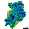
 Map data Map data | Reconstruction of human pre-40S particle purified by using a HASt-tagged version of the ribosome biogenesis factor LTV1 as bait. the map has been filtered and masked using the RELION1.3 postprocess option. | |||||||||
 Sample Sample |
| |||||||||
 Keywords Keywords |  ribosome / small subunit precursor / human cells ribosome / small subunit precursor / human cells | |||||||||
| Biological species |  Homo sapiens (human) Homo sapiens (human) | |||||||||
| Method | single particle reconstruction / cryo EM / Resolution: 19.4 Å | |||||||||
 Authors Authors | Larburu N / Montellese C / O'Donohue M-F / Kutay U / Gleizes P-E / Plisson-Chastang C | |||||||||
 Citation Citation | Journal: Nucleic Acids Res / Year: 2016 Title: Structure of a human pre-40S particle points to a role for RACK1 in the final steps of 18S rRNA processing. Authors: Natacha Larburu / Christian Montellese / Marie-Françoise O'Donohue / Ulrike Kutay / Pierre-Emmanuel Gleizes / Célia Plisson-Chastang /   Abstract: Synthesis of ribosomal subunits in eukaryotes is a complex and tightly regulated process that has been mostly characterized in yeast. The discovery of a growing number of diseases linked to defects ...Synthesis of ribosomal subunits in eukaryotes is a complex and tightly regulated process that has been mostly characterized in yeast. The discovery of a growing number of diseases linked to defects in ribosome biogenesis calls for a deeper understanding of these mechanisms and of the specificities of human ribosome maturation. We present the 19 Å resolution cryo-EM reconstruction of a cytoplasmic precursor to the human small ribosomal subunit, purified by using the tagged ribosome biogenesis factor LTV1 as bait. Compared to yeast pre-40S particles, this first three-dimensional structure of a human 40S subunit precursor shows noticeable differences with respect to the position of ribosome biogenesis factors and uncovers the early deposition of the ribosomal protein RACK1 during subunit maturation. Consistently, RACK1 is required for efficient processing of the 18S rRNA 3'-end, which might be related to its role in translation initiation. This first structural analysis of a human pre-ribosomal particle sets the grounds for high-resolution studies of conformational transitions accompanying ribosomal subunit maturation. | |||||||||
| History |
|
- Structure visualization
| Movie |
 Movie viewer Movie viewer |
|---|---|
| Structure viewer | EM map: SurfViewMolmilJmol/JSmol |
| Supplemental images |

- Downloads & links
-EMDB archive
| Map data | emd_3300.map.gz | 3.5 MB | EMDB map data format | |
|---|---|---|---|---|
| Header (meta data) | emd-3300-v30.xmlemd-3300.xml | 9.8 KB 9.8 KB | Display Display | EMDB header |
| FSC (resolution estimation) | emd_3300_fsc.xml | 6.6 KB | Display | FSC data file |
| Images | emd_3300.tif | 1.3 MB | ||
| Archive directory |  http://ftp.pdbj.org/pub/emdb/structures/EMD-3300ftp://ftp.pdbj.org/pub/emdb/structures/EMD-3300 http://ftp.pdbj.org/pub/emdb/structures/EMD-3300ftp://ftp.pdbj.org/pub/emdb/structures/EMD-3300 | HTTPS FTP |
-Related structure data
| Similar structure data |
|---|

-Links
| EMDB pages | EMDB (EBI/PDBe) / EMDataResource |
|---|---|
| Related items in Molecule of the Month |

-Map
| File | Download / File: emd_3300.map.gz / Format: CCP4 / Size: 26.4 MB / Type: IMAGE STORED AS FLOATING POINT NUMBER (4 BYTES) | ||||||||||||||||||||||||||||||||||||||||||||||||||||||||||||||||||||
|---|---|---|---|---|---|---|---|---|---|---|---|---|---|---|---|---|---|---|---|---|---|---|---|---|---|---|---|---|---|---|---|---|---|---|---|---|---|---|---|---|---|---|---|---|---|---|---|---|---|---|---|---|---|---|---|---|---|---|---|---|---|---|---|---|---|---|---|---|---|
| Annotation | Reconstruction of human pre-40S particle purified by using a HASt-tagged version of the ribosome biogenesis factor LTV1 as bait. the map has been filtered and masked using the RELION1.3 postprocess option. | ||||||||||||||||||||||||||||||||||||||||||||||||||||||||||||||||||||
| Voxel size | X=Y=Z: 2.27 Å | ||||||||||||||||||||||||||||||||||||||||||||||||||||||||||||||||||||
| Density |
| ||||||||||||||||||||||||||||||||||||||||||||||||||||||||||||||||||||
| Symmetry | Space group: 1 | ||||||||||||||||||||||||||||||||||||||||||||||||||||||||||||||||||||
| Details | EMDB XML:
CCP4 map header:
| ||||||||||||||||||||||||||||||||||||||||||||||||||||||||||||||||||||
-Supplemental data
- Sample components
-Entire : pre-40S particles purified from human cells by using the tagged r...
| Entire | Name: pre-40S particles purified from human cells by using the tagged ribosome biogenesis factor HASt_LTV1 as bait. |
|---|---|
| Components |
|
-Supramolecule #1000: pre-40S particles purified from human cells by using the tagged r...
| Supramolecule | Name: pre-40S particles purified from human cells by using the tagged ribosome biogenesis factor HASt_LTV1 as bait. type: sample / ID: 1000 / Oligomeric state: monomer / Number unique components: 1 |
|---|---|
| Molecular weight | Theoretical: 1.5 MDa |
-Supramolecule #1: cytoplasmic precursor of the human small ribosomal subunit
| Supramolecule | Name: cytoplasmic precursor of the human small ribosomal subunit type: complex / ID: 1 / Name.synonym: pre-40S particle / Recombinant expression: No / Ribosome-details: ribosome-eukaryote: SSU 40S |
|---|---|
| Source (natural) | Organism: Homo sapiens (human) / synonym: Human / Tissue: Embryonic Kidney / Cell: HEK 293 |
| Molecular weight | Theoretical: 1.5 MDa |
-Experimental details
-Structure determination
| Method | cryo EM |
|---|---|
 Processing Processing | single particle reconstruction |
| Aggregation state | particle |
-Sample preparation
| Concentration | 0.02 mg/mL |
|---|---|
| Buffer | pH: 7.6 / Details: 10 mM Tris-HCl, 100 mM KCl, 2 mM MgCl2 |
| Grid | Details: Glow discharged Quantifoil R2/1 on Cu 300 Mesh grids |
| Vitrification | Cryogen name: ETHANE / Chamber humidity: 95 % / Chamber temperature: 293 K / Instrument: LEICA EM GP / Method: 1.8 - 2.1'' blot before plunging |
- Electron microscopy
| Microscope | FEI TITAN KRIOS |
|---|---|
| Electron beam | Acceleration voltage: 300 kV / Electron source: FIELD EMISSION GUN |
| Electron optics | Illumination mode: FLOOD BEAM / Imaging mode: BRIGHT FIELDBright-field microscopy / Cs: 0.02 mm / Nominal defocus max: 3.6 µm / Nominal defocus min: 1.2 µm / Nominal magnification: 59000 |
| Sample stage | Specimen holder model: FEI TITAN KRIOS AUTOGRID HOLDER |
| Temperature | Average: 100 K |
| Date | Jan 23, 2014 |
| Image recording | Category: CCD / Film or detector model: FEI FALCON II (4k x 4k) / Number real images: 3812 / Average electron dose: 30 e/Å2 Details: Every image has been acquired in 1s by the direct electron detector (no movies recorded) |
| Tilt angle min | 0 |
| Tilt angle max | 0 |
| Experimental equipment |  Model: Titan Krios / Image courtesy: FEI Company |
-Image processing
| CTF correction | Details: CTFFIND3 |
|---|---|
| Final reconstruction | Applied symmetry - Point group: C1 (asymmetric) / Resolution.type: BY AUTHOR / Resolution: 19.4 Å / Resolution method: OTHER / Software - Name: RELION1.3 / Number images used: 54436 |
| Details | Particles were picked with e2boxer.py fro EMAN2 Reconstruction was carried out with Relion 1.3 |
| FSC plot (resolution estimation) |  |
-Atomic model buiding 1
| Initial model | PDB ID: |
|---|---|
| Software | Name: Chimera |
| Details | The 40S components of pdb model 4V6X were fitted into the pre-40S map by rigid body docking. |
| Refinement | Space: REAL / Protocol: RIGID BODY FIT |