 Movie
Movie Controller

[English] 日本語
 Yorodumi
- EMDB-3281: Antigenic and cryo-electron microscopy structure analysis of a ch... -
+ Open data

- Basic information
| Entry | Database: EMDB / ID: EMD-3281 | |||||||||
|---|---|---|---|---|---|---|---|---|---|---|
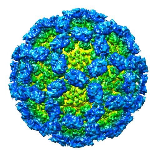
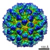
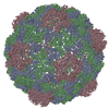
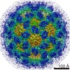
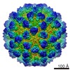
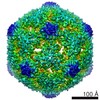
| Title | Antigenic and cryo-electron microscopy structure analysis of a chimeric sapovirus capsid | |||||||||
 Map data Map data | Chimeric human sapovirus capsid | |||||||||
 Sample Sample |
| |||||||||
 Keywords Keywords | non-envelope virus / VLP / calicivirus | |||||||||
| Biological species |  Sapovirus Sapovirus | |||||||||
| Method | single particle reconstruction / cryo EM / negative staining / Resolution: 8.5 Å | |||||||||
 Authors Authors | Miyazaki N / Taylor DW / Hansman GS / Murata K | |||||||||
 Citation Citation | Journal: J Virol / Year: 2015 Title: Antigenic and Cryo-Electron Microscopy Structure Analysis of a Chimeric Sapovirus Capsid. Authors: Naoyuki Miyazaki / David W Taylor / Grant S Hansman / Kazuyoshi Murata /    Abstract: The capsid protein (VP1) of all caliciviruses forms an icosahedral particle with two principal domains, shell (S) and protruding (P) domains, which are connected via a flexible hinge region. The S ...The capsid protein (VP1) of all caliciviruses forms an icosahedral particle with two principal domains, shell (S) and protruding (P) domains, which are connected via a flexible hinge region. The S domain forms a scaffold surrounding the nucleic acid, while the P domains form a homodimer that interacts with receptors. The P domain is further subdivided into two subdomains, termed P1 and P2. The P2 subdomain is likely an insertion in the P1 subdomain; consequently, the P domain is divided into the P1-1, P2, and P1-2 subdomains. In order to investigate capsid antigenicity, N-terminal (N-term)/S/P1-1 and P2/P1-2 were switched between two sapovirus genotypes GI.1 and GI.5. The chimeric VP1 constructs were expressed in insect cells and were shown to self-assemble into virus-like particles (VLPs) morphologically similar to the parental VLPs. Interestingly, the chimeric VLPs had higher levels of cross-reactivities to heterogeneous antisera than the parental VLPs. In order to better understand the antigenicity from a structural perspective, we determined an intermediate-resolution (8.5-Å) cryo-electron microscopy (cryo-EM) structure of a chimeric VLP and developed a VP1 homology model. The cryo-EM structure revealed that the P domain dimers were raised slightly (∼5 Å) above the S domain. The VP1 homology model allowed us predict the S domain (67-229) and P1-1 (229-280), P2 (281-447), and P1-2 (448-567) subdomains. Our results suggested that the raised P dimers might expose immunoreactive S/P1-1 subdomain epitopes. Consequently, the higher levels of cross-reactivities with the chimeric VLPs resulted from a combination of GI.1 and GI.5 epitopes. IMPORTANCE: We developed sapovirus chimeric VP1 constructs and produced the chimeric VLPs in insect cells. We found that both chimeric VLPs had a higher level of cross-reactivity against ...IMPORTANCE: We developed sapovirus chimeric VP1 constructs and produced the chimeric VLPs in insect cells. We found that both chimeric VLPs had a higher level of cross-reactivity against heterogeneous VLP antisera than the parental VLPs. The cryo-EM structure of one chimeric VLP (Yokote/Mc114) was solved to 8.5-Å resolution. A homology model of the VP1 indicated for the first time the putative S and P (P1-1, P2, and P1-2) domains. The overall structure of Yokote/Mc114 contained features common among other caliciviruses. We showed that the P2 subdomain was mainly involved in the homodimeric interface, whereas a large gap between the P1 subdomains had fewer interactions. | |||||||||
| History |
|
- Structure visualization
| Movie |
 Movie viewer Movie viewer |
|---|---|
| Structure viewer | EM map: SurfViewMolmilJmol/JSmol |
| Supplemental images |
- Downloads & links
-EMDB archive
| Map data | emd_3281.map.gz | 115.7 MB | EMDB map data format | |
|---|---|---|---|---|
| Header (meta data) | emd-3281-v30.xmlemd-3281.xml | 9.3 KB 9.3 KB | Display Display | EMDB header |
| Images |  EMD-3281.png EMD-3281.png | 328.8 KB | ||
| Archive directory |  http://ftp.pdbj.org/pub/emdb/structures/EMD-3281ftp://ftp.pdbj.org/pub/emdb/structures/EMD-3281 http://ftp.pdbj.org/pub/emdb/structures/EMD-3281ftp://ftp.pdbj.org/pub/emdb/structures/EMD-3281 | HTTPS FTP |
-Validation report
| Summary document | emd_3281_validation.pdf.gz | 312.4 KB | Display | EMDB validaton report |
|---|---|---|---|---|
| Full document | emd_3281_full_validation.pdf.gz | 311.5 KB | Display | |
| Data in XML | emd_3281_validation.xml.gz | 7.6 KB | Display | |
| Arichive directory | https://ftp.pdbj.org/pub/emdb/validation_reports/EMD-3281ftp://ftp.pdbj.org/pub/emdb/validation_reports/EMD-3281 | HTTPS FTP |
-Related structure data
| Similar structure data |
|---|
-Links
| EMDB pages | EMDB (EBI/PDBe) / EMDataResource |
|---|
-Map
| File | Download / File: emd_3281.map.gz / Format: CCP4 / Size: 127.9 MB / Type: IMAGE STORED AS FLOATING POINT NUMBER (4 BYTES) | ||||||||||||||||||||||||||||||||||||||||||||||||||||||||||||
|---|---|---|---|---|---|---|---|---|---|---|---|---|---|---|---|---|---|---|---|---|---|---|---|---|---|---|---|---|---|---|---|---|---|---|---|---|---|---|---|---|---|---|---|---|---|---|---|---|---|---|---|---|---|---|---|---|---|---|---|---|---|
| Annotation | Chimeric human sapovirus capsid | ||||||||||||||||||||||||||||||||||||||||||||||||||||||||||||
| Voxel size | X=Y=Z: 1.62 Å | ||||||||||||||||||||||||||||||||||||||||||||||||||||||||||||
| Density |
| ||||||||||||||||||||||||||||||||||||||||||||||||||||||||||||
| Symmetry | Space group: 1 | ||||||||||||||||||||||||||||||||||||||||||||||||||||||||||||
| Details | EMDB XML:
CCP4 map header:
| ||||||||||||||||||||||||||||||||||||||||||||||||||||||||||||
-Supplemental data
- Sample components
-Entire : Chimera human sapovirus capsid
| Entire | Name: Chimera human sapovirus capsid |
|---|---|
| Components |
|
-Supramolecule #1000: Chimera human sapovirus capsid
| Supramolecule | Name: Chimera human sapovirus capsid / type: sample / ID: 1000 / Oligomeric state: icosahedral / Number unique components: 1 |
|---|---|
| Molecular weight | Experimental: 10 MDa / Theoretical: 10 MDa |
-Supramolecule #1: Sapovirus
| Supramolecule | Name: Sapovirus / type: virus / ID: 1 / Name.synonym: human sapovirus / NCBI-ID: 95341 / Sci species name: Sapovirus / Virus type: VIRUS-LIKE PARTICLE / Virus isolate: OTHER / Virus enveloped: No / Virus empty: Yes / Syn species name: human sapovirus |
|---|---|
| Host (natural) | Organism:  Homo sapiens (human) / synonym: VERTEBRATES Homo sapiens (human) / synonym: VERTEBRATES |
| Host system | Organism: unidentified baculovirus |
| Molecular weight | Experimental: 10 MDa / Theoretical: 10 MDa |
| Virus shell | Shell ID: 1 / Diameter: 38 Å / T number (triangulation number): 3 |
-Experimental details
-Structure determination
| Method | negative staining, cryo EM |
|---|---|
 Processing Processing | single particle reconstruction |
| Aggregation state | particle |
-Sample preparation
| Staining | Type: NEGATIVE / Details: ice-embedding |
|---|---|
| Grid | Details: quantifoil R1.2/1.3 |
| Vitrification | Cryogen name: ETHANE / Chamber humidity: 95 % / Chamber temperature: 120 K / Instrument: FEI VITROBOT MARK III |
- Electron microscopy
| Microscope | JEOL 2200FS |
|---|---|
| Temperature | Min: 90 K / Max: 105 K / Average: 100 K |
| Specialist optics | Energy filter - Name: Omega-type / Energy filter - Lower energy threshold: 0.0 eV / Energy filter - Upper energy threshold: 15.0 eV |
| Date | Jul 15, 2012 |
| Image recording | Category: CCD / Film or detector model: TVIPS TEMCAM-F415 (4k x 4k) / Digitization - Sampling interval: 15 µm / Number real images: 80 / Average electron dose: 20 e/Å2 / Od range: 5 / Bits/pixel: 16 |
| Electron beam | Acceleration voltage: 200 kV / Electron source:  FIELD EMISSION GUN FIELD EMISSION GUN |
| Electron optics | Calibrated magnification: 91463 / Illumination mode: FLOOD BEAM / Imaging mode: BRIGHT FIELD / Cs: 4.2 mm / Nominal defocus max: 3.0 µm / Nominal defocus min: 1.0 µm / Nominal magnification: 60000 |
| Sample stage | Specimen holder model: GATAN LIQUID NITROGEN |
-Image processing
| CTF correction | Details: Each particle |
|---|---|
| Final reconstruction | Applied symmetry - Point group: I (icosahedral) / Algorithm: OTHER / Resolution.type: BY AUTHOR / Resolution: 8.5 Å / Resolution method: OTHER / Software - Name: EMAN2 / Number images used: 6000 |
