 Movie
Movie Controller

[English] 日本語
 Yorodumi
- EMDB-32742: Structures of Omicron Spike complexes illuminate broad-spectrum n... -
+ Open data

- Basic information
| Entry | 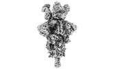 | |||||||||
|---|---|---|---|---|---|---|---|---|---|---|
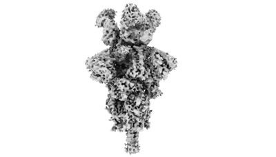
| Title | Structures of Omicron Spike complexes illuminate broad-spectrum neutralizing antibody development | |||||||||
 Map data Map data | ||||||||||
 Sample Sample |
| |||||||||
| Function / homology |  Function and homology information Function and homology informationMaturation of spike protein / viral translation / Translation of Structural Proteins / Virion Assembly and Release / host cell surface / host extracellular space / suppression by virus of host tetherin activity / Induction of Cell-Cell Fusion / structural constituent of virion / entry receptor-mediated virion attachment to host cell ...Maturation of spike protein / viral translation / Translation of Structural Proteins / Virion Assembly and Release / host cell surface / host extracellular space / suppression by virus of host tetherin activity / Induction of Cell-Cell Fusion / structural constituent of virion / entry receptor-mediated virion attachment to host cell / host cell endoplasmic reticulum-Golgi intermediate compartment membrane / receptor-mediated endocytosis of virus by host cell / Attachment and Entry /  membrane fusion / positive regulation of viral entry into host cell / receptor-mediated virion attachment to host cell / receptor ligand activity / host cell surface receptor binding / fusion of virus membrane with host plasma membrane / fusion of virus membrane with host endosome membrane / viral envelope / symbiont-mediated suppression of host type I interferon-mediated signaling pathway / virion attachment to host cell / SARS-CoV-2 activates/modulates innate and adaptive immune responses / host cell plasma membrane / virion membrane / membrane / identical protein binding / plasma membrane membrane fusion / positive regulation of viral entry into host cell / receptor-mediated virion attachment to host cell / receptor ligand activity / host cell surface receptor binding / fusion of virus membrane with host plasma membrane / fusion of virus membrane with host endosome membrane / viral envelope / symbiont-mediated suppression of host type I interferon-mediated signaling pathway / virion attachment to host cell / SARS-CoV-2 activates/modulates innate and adaptive immune responses / host cell plasma membrane / virion membrane / membrane / identical protein binding / plasma membraneSimilarity search - Function | |||||||||
| Biological species |   Severe acute respiratory syndrome coronavirus 2 / Severe acute respiratory syndrome coronavirus 2 /  Homo sapiens (human) Homo sapiens (human) | |||||||||
| Method | single particle reconstruction / cryo EM / Resolution: 3.6 Å | |||||||||
 Authors Authors | Guo H / Gao Y / Ji X / Yang H | |||||||||
| Funding support |  China, 1 items China, 1 items
| |||||||||
 Citation Citation | Journal: Cell Rep / Year: 2022 Title: Structures of Omicron spike complexes and implications for neutralizing antibody development. Authors: Hangtian Guo / Yan Gao / Tinghan Li / Tingting Li / Yuchi Lu / Le Zheng / Yue Liu / Tingting Yang / Feiyang Luo / Shuyi Song / Wei Wang / Xiuna Yang / Henry C Nguyen / Hongkai Zhang / Ailong ...Authors: Hangtian Guo / Yan Gao / Tinghan Li / Tingting Li / Yuchi Lu / Le Zheng / Yue Liu / Tingting Yang / Feiyang Luo / Shuyi Song / Wei Wang / Xiuna Yang / Henry C Nguyen / Hongkai Zhang / Ailong Huang / Aishun Jin / Haitao Yang / Zihe Rao / Xiaoyun Ji /  Abstract: The emergence of the SARS-CoV-2 Omicron variant is dominant in many countries worldwide. The high number of spike mutations is responsible for the broad immune evasion from existing vaccines and ...The emergence of the SARS-CoV-2 Omicron variant is dominant in many countries worldwide. The high number of spike mutations is responsible for the broad immune evasion from existing vaccines and antibody drugs. To understand this, we first present the cryo-electron microscopy structure of ACE2-bound SARS-CoV-2 Omicron spike. Comparison to previous spike antibody structures explains how Omicron escapes these therapeutics. Secondly, we report structures of Omicron, Delta, and wild-type spikes bound to a patient-derived Fab antibody fragment (510A5), which provides direct evidence where antibody binding is greatly attenuated by the Omicron mutations, freeing spike to bind ACE2. Together with biochemical binding and 510A5 neutralization assays, our work establishes principles of binding required for neutralization and clearly illustrates how the mutations lead to antibody evasion yet retain strong ACE2 interactions. Structural information on spike with both bound and unbound antibodies collectively elucidates potential strategies for generation of therapeutic antibodies. | |||||||||
| History |
|
- Structure visualization
| Supplemental images |
|---|
- Downloads & links
-EMDB archive
| Map data | emd_32742.map.gz | 483.6 MB | EMDB map data format | |
|---|---|---|---|---|
| Header (meta data) | emd-32742-v30.xmlemd-32742.xml | 14.9 KB 14.9 KB | Display Display | EMDB header |
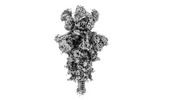
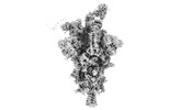
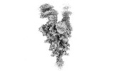
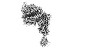
| Images |  emd_32742.png emd_32742.png | 35.9 KB | ||
| Archive directory |  http://ftp.pdbj.org/pub/emdb/structures/EMD-32742ftp://ftp.pdbj.org/pub/emdb/structures/EMD-32742 http://ftp.pdbj.org/pub/emdb/structures/EMD-32742ftp://ftp.pdbj.org/pub/emdb/structures/EMD-32742 | HTTPS FTP |
-Related structure data
| Related structure data | 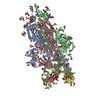 7ws3MC 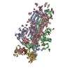 7ws0C 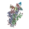 7ws1C 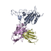 7ws2C 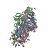 7ws4C 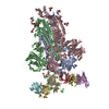 7ws5C 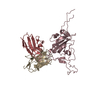 7ws6C 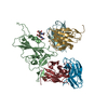 7ws7C 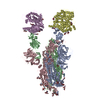 7ws8C 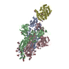 7ws9C 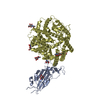 7wsaC M: atomic model generated by this map C: citing same article ( |
|---|---|
| Similar structure data |


-Links
| EMDB pages | EMDB (EBI/PDBe) / EMDataResource |
|---|---|
| Related items in Molecule of the Month |


-Map
| File | Download / File: emd_32742.map.gz / Format: CCP4 / Size: 512 MB / Type: IMAGE STORED AS FLOATING POINT NUMBER (4 BYTES) | ||||||||||||||||||||
|---|---|---|---|---|---|---|---|---|---|---|---|---|---|---|---|---|---|---|---|---|---|
| Voxel size | X=Y=Z: 0.82 Å | ||||||||||||||||||||
| Density |
| ||||||||||||||||||||
| Symmetry | Space group: 1 | ||||||||||||||||||||
| Details | EMDB XML:
|
-Supplemental data
- Sample components
-Entire : SARS-CoV-2 Delta variant spike glycoprotein complex with 510A5 Fab
| Entire | Name: SARS-CoV-2 Delta variant spike glycoprotein complex with 510A5 Fab |
|---|---|
| Components |
|
-Supramolecule #1: SARS-CoV-2 Delta variant spike glycoprotein complex with 510A5 Fab
| Supramolecule | Name: SARS-CoV-2 Delta variant spike glycoprotein complex with 510A5 Fab type: complex / Chimera: Yes / ID: 1 / Parent: 0 / Macromolecule list: #1-#3 |
|---|---|
| Source (natural) | Organism: Severe acute respiratory syndrome coronavirus 2 |
-Supramolecule #2: Delta spike glycoprotein
| Supramolecule | Name: Delta spike glycoprotein / type: complex / Chimera: Yes / ID: 2 / Parent: 1 / Macromolecule list: #1 / Details: SARS-CoV-2 Delta variant Spike protein ectodomain |
|---|---|
| Source (natural) | Organism: Homo sapiens (human) |
| Recombinant expression | Organism: Homo sapiens (human) / Recombinant cell: HEK293 |
-Supramolecule #3: Fab
| Supramolecule | Name: Fab / type: complex / Chimera: Yes / ID: 3 / Parent: 1 / Macromolecule list: #2-#3 Details: Fab fragment generated by proteolytic cleavage of IgG antibody |
|---|
-Macromolecule #1: Spike glycoprotein
| Macromolecule | Name: Spike glycoprotein / type: protein_or_peptide / ID: 1 / Number of copies: 3 / Enantiomer: LEVO |
|---|---|
| Source (natural) | Organism: Severe acute respiratory syndrome coronavirus 2 |
| Molecular weight | Theoretical: 142.374406 KDa |
| Recombinant expression | Organism: Homo sapiens (human) |
| Sequence | String: MFVFLVLLPL VSSQCVNLRT RTQLPPAYTN SFTRGVYYPD KVFRSSVLHS TQDLFLPFFS NVTWFHAIHV SGTNGTKRFD NPVLPFNDG VYFASIEKSN IIRGWIFGTT LDSKTQSLLI VNNATNVVIK VCEFQFCNDP FLDVYYHKNN KSWMESECGV Y SSANNCTF ...String: MFVFLVLLPL VSSQCVNLRT RTQLPPAYTN SFTRGVYYPD KVFRSSVLHS TQDLFLPFFS NVTWFHAIHV SGTNGTKRFD NPVLPFNDG VYFASIEKSN IIRGWIFGTT LDSKTQSLLI VNNATNVVIK VCEFQFCNDP FLDVYYHKNN KSWMESECGV Y SSANNCTF EYVSQPFLMD LEGKQGNFKN LREFVFKNID GYFKIYSKHT PINLVRDLPQ GFSALEPLVD LPIGINITRF QT LLALHRS YLTPGDSSSG WTAGAAAYYV GYLQPRTFLL KYNENGTITD AVDCALDPLS ETKCTLKSFT VEKGIYQTSN FRV QPTESI VRFPNITNLC PFGEVFNATR FASVYAWNRK RISNCVADYS VLYNSASFST FKCYGVSPTK LNDLCFTNVY ADSF VIRGD EVRQIAPGQT GKIADYNYKL PDDFTGCVIA WNSNNLDSKV GGNYNYRYRL FRKSNLKPFE RDISTEIYQA GSKPC NGVE GFNCYFPLQS YGFQPTNGVG YQPYRVVVLS FELLHAPATV CGPKKSTNLV KNKCVNFNFN GLTGTGVLTE SNKKFL PFQ QFGRDIADTT DAVRDPQTLE ILDITPCSFG GVSVITPGTN TSNQVAVLYQ GVNCTEVPVA IHADQLTPTW RVYSTGS NV FQTRAGCLIG AEHVNNSYEC DIPIGAGICA SYQTQTNSRG SASSVASQSI IAYTMSLGAE NSVAYSNNSI AIPTNFTI S VTTEILPVSM TKTSVDCTMY ICGDSTECSN LLLQYGSFCT QLNRALTGIA VEQDKNTQEV FAQVKQIYKT PPIKDFGGF NFSQILPDPS KPSKRSFIED LLFNKVTLAD AGFIKQYGDC LGDIAARDLI CAQKFNGLTV LPPLLTDEMI AQYTSALLAG TITSGWTFG AGAALQIPFA MQMAYRFNGI GVTQNVLYEN QKLIANQFNS AIGKIQDSLS STASALGKLQ NVVNQNAQAL N TLVKQLSS NFGAISSVLN DILSRLDPPE AEVQIDRLIT GRLQSLQTYV TQQLIRAAEI RASANLAATK MSECVLGQSK RV DFCGKGY HLMSFPQSAP HGVVFLHVTY VPAQEKNFTT APAICHDGKA HFPREGVFVS NGTHWFVTQR NFYEPQIITT DNT FVSGNC DVVIGIVNNT VYDPLQPELD SFKEELDKYF KNHTSPDVDL GDISGINASV VNIQKEIDRL NEVAKNLNES LIDL QELGK YEQGSGYIPE APRDGQAYVR KDGEWVFLST FLSGLEVLFQ GPGGWSHPQF EKGGGSGGGS GGSAWSHPQF EKGGS HHHH HHHH |
-Macromolecule #2: 510A5 light chain
| Macromolecule | Name: 510A5 light chain / type: protein_or_peptide / ID: 2 / Number of copies: 3 / Enantiomer: LEVO |
|---|---|
| Source (natural) | Organism: Homo sapiens (human) |
| Molecular weight | Theoretical: 11.680938 KDa |
| Recombinant expression | Organism: Homo sapiens (human) |
| Sequence | String: DIQMTQSPSS LSASVGDRVT ITCRASQSIS SYLNWFQHKP GKAPKLLIYG ASSLQSGVPS RFSGSGSGTD FTLTISSLQP EDFATYYCQ QSYSTPPYTF GQGTKLEIK |
-Macromolecule #3: 510A5 heavy chain
| Macromolecule | Name: 510A5 heavy chain / type: protein_or_peptide / ID: 3 / Number of copies: 3 / Enantiomer: LEVO |
|---|---|
| Source (natural) | Organism: Homo sapiens (human) |
| Molecular weight | Theoretical: 13.586025 KDa |
| Recombinant expression | Organism: Homo sapiens (human) |
| Sequence | String: EVQLVESGGG LVQPGRSLRL SCAASGFTFD DYAMHWVRQA PGKGLEWVSG ISWNSDSIDY ADSVKGRFTI SRDNAKNSLY LQMNSLRAE DTALYYCAKD RGYEILTPAS FDYWGQGTLV TVSS |
-Macromolecule #5: 2-acetamido-2-deoxy-beta-D-glucopyranose
| Macromolecule | Name: 2-acetamido-2-deoxy-beta-D-glucopyranose / type: ligand / ID: 5 / Number of copies: 24 / Formula: NAG |
|---|---|
| Molecular weight | Theoretical: 221.208 Da |
| Chemical component information |  ChemComp-NAG: |
-Experimental details
-Structure determination
| Method | cryo EM |
|---|---|
 Processing Processing | single particle reconstruction |
| Aggregation state | particle |
-Sample preparation
| Buffer | pH: 7.4 |
|---|---|
| Vitrification | Cryogen name: ETHANE |
- Electron microscopy
| Microscope | FEI TITAN KRIOS |
|---|---|
| Electron beam | Acceleration voltage: 300 kV / Electron source: FIELD EMISSION GUN |
| Electron optics | Illumination mode: SPOT SCAN / Imaging mode: BRIGHT FIELDBright-field microscopy / Nominal defocus max: 2.8000000000000003 µm / Nominal defocus min: 1.2 µm |
| Image recording | Film or detector model: GATAN K3 (6k x 4k) / Average electron dose: 60.0 e/Å2 |
| Experimental equipment |  Model: Titan Krios / Image courtesy: FEI Company |
-Image processing
| Initial angle assignment | Type: COMMON LINE |
|---|---|
| Final angle assignment | Type: COMMON LINE |
| Final reconstruction | Resolution.type: BY AUTHOR / Resolution: 3.6 Å / Resolution method: FSC 0.143 CUT-OFF / Number images used: 141036 |
