 Movie
Movie Controller


+ Open data

- Basic information
| Entry |  | |||||||||
|---|---|---|---|---|---|---|---|---|---|---|
| Title | Structure of Apoferritin | |||||||||
 Map data Map data | the reconstruction of full frame | |||||||||
 Sample Sample |
| |||||||||
 Keywords Keywords |  complex / TRANSPORT PROTEIN / OXIDOREDUCTASE complex / TRANSPORT PROTEIN / OXIDOREDUCTASE | |||||||||
| Function / homology |  Function and homology information Function and homology informationiron ion sequestering activity / : / autolysosome / Scavenging by Class A Receptors / Golgi Associated Vesicle Biogenesis / ferroxidase / intracellular sequestering of iron ion / ferroxidase activity / negative regulation of fibroblast proliferation / ferric iron binding ...iron ion sequestering activity / : / autolysosome / Scavenging by Class A Receptors / Golgi Associated Vesicle Biogenesis / ferroxidase / intracellular sequestering of iron ion / ferroxidase activity / negative regulation of fibroblast proliferation / ferric iron binding / ferrous iron binding / Iron uptake and transport / tertiary granule lumen / iron ion transport / intracellular iron ion homeostasis / ficolin-1-rich granule lumen / immune response / iron ion binding / negative regulation of cell population proliferation / Neutrophil degranulation / extracellular exosome / extracellular region / identical protein binding / nucleus / cytosol / cytoplasmSimilarity search - Function | |||||||||
| Biological species |  Homo sapiens (human) Homo sapiens (human) | |||||||||
| Method | single particle reconstruction / cryo EM / Resolution: 1.89 Å | |||||||||
 Authors Authors | Zhang X / Wu C / Shi H | |||||||||
| Funding support | 1 items
| |||||||||
 Citation Citation | Journal: QRB Discov / Year: 2021 Title: Low-cooling-rate freezing in biomolecular cryo-electron microscopy for recovery of initial frames. Authors: Chunling Wu / Huigang Shi / Dongjie Zhu / Kelong Fan / Xinzheng Zhang /  Abstract: When biological samples are first exposed to electrons in cryo-electron microcopy (cryo-EM), proteins exhibit a rapid 'burst' phase of beam-induced motion that cannot be corrected with software. This ...When biological samples are first exposed to electrons in cryo-electron microcopy (cryo-EM), proteins exhibit a rapid 'burst' phase of beam-induced motion that cannot be corrected with software. This lowers the quality of the initial frames, which are the least damaged by the electrons. Hence, they are commonly excluded or down-weighted during data processing, reducing the undamaged signal and the resolution in the reconstruction. By decreasing the cooling rate during sample preparation, either with a cooling-rate gradient or by increasing the freezing temperature, we show that the quality of the initial frames for various protein and virus samples can be recovered. Incorporation of the initial frames in the reconstruction increases the resolution by an amount equivalent to using ~60% more data. Moreover, these frames preserve the high-quality cryo-EM densities of radiation-sensitive residues, which is often damaged or very weak in canonical three-dimensional reconstruction. The improved freezing conditions can be easily achieved using existing devices and enhance the overall quality of cryo-EM structures. | |||||||||
| History |
|
- Structure visualization
| Supplemental images |
|---|

- Downloads & links
-EMDB archive
| Map data | emd_31736.map.gz | 228.3 MB | EMDB map data format | |
|---|---|---|---|---|
| Header (meta data) | emd-31736-v30.xmlemd-31736.xml | 29.4 KB 29.4 KB | Display Display | EMDB header |
| Images |  emd_31736.png emd_31736.png | 228.4 KB | ||
| Others | emd_31736_additional_1.map.gzemd_31736_additional_10.map.gzemd_31736_additional_2.map.gzemd_31736_additional_3.map.gzemd_31736_additional_4.map.gzemd_31736_additional_5.map.gzemd_31736_additional_6.map.gzemd_31736_additional_7.map.gzemd_31736_additional_8.map.gzemd_31736_additional_9.map.gz | 218.4 MB 218.5 MB 218.8 MB 218.9 MB 218.7 MB 218.9 MB 218.9 MB 219 MB 218.6 MB 218.3 MB | ||
| Archive directory |  http://ftp.pdbj.org/pub/emdb/structures/EMD-31736ftp://ftp.pdbj.org/pub/emdb/structures/EMD-31736 http://ftp.pdbj.org/pub/emdb/structures/EMD-31736ftp://ftp.pdbj.org/pub/emdb/structures/EMD-31736 | HTTPS FTP |
-Related structure data
| Related structure data | 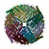 7v66MC M: atomic model generated by this map C: citing same article ( |
|---|---|
| Similar structure data |
-Links
| EMDB pages | EMDB (EBI/PDBe) / EMDataResource |
|---|---|
| Related items in Molecule of the Month |


-Map
| File | Download / File: emd_31736.map.gz / Format: CCP4 / Size: 244.1 MB / Type: IMAGE STORED AS FLOATING POINT NUMBER (4 BYTES) | ||||||||||||||||||||
|---|---|---|---|---|---|---|---|---|---|---|---|---|---|---|---|---|---|---|---|---|---|
| Annotation | the reconstruction of full frame | ||||||||||||||||||||
| Voxel size | X=Y=Z: 0.515 Å | ||||||||||||||||||||
| Density |
| ||||||||||||||||||||
| Symmetry | Space group: 1 | ||||||||||||||||||||
| Details | EMDB XML:
|
-Supplemental data
+Additional map: the eighth frame of per-frame reconstruction
 Z
Z Y
Y X
X


+Additional map: the tenth frame of per-frame reconstruction


+Additional map: the seventh frame of per-frame reconstruction


+Additional map: the second frame of per-frame reconstruction


+Additional map: the first frame of per-frame reconstruction


+Additional map: the third frame of per-frame reconstruction


+Additional map: the fifth frame of per-frame reconstruction


+Additional map: the fourth frame of per-frame reconstruction


+Additional map: the sixth frame of per-frame reconstruction


+Additional map: the ninth frame of per-frame reconstruction


- Sample components
-Entire : Human apoferritin
| Entire | Name: Human apoferritinFerritin |
|---|---|
| Components |
|
-Supramolecule #1: Human apoferritin
| Supramolecule | Name: Human apoferritin / type: complex / ID: 1 / Parent: 0 / Macromolecule list: all |
|---|---|
| Source (natural) | Organism: Homo sapiens (human) |
| Molecular weight | Theoretical: 440 KDa |
-Macromolecule #1: Ferritin heavy chain
| Macromolecule | Name: Ferritin heavy chain / type: protein_or_peptide / ID: 1 / Number of copies: 24 / Enantiomer: LEVO / EC number: ferroxidase |
|---|---|
| Source (natural) | Organism: Homo sapiens (human) |
| Molecular weight | Theoretical: 20.116547 KDa |
| Recombinant expression | Organism:  Escherichia coli (E. coli) Escherichia coli (E. coli) |
| Sequence | String: TSQVRQNYHQ DSEAAINRQI NLELYASYVY LSMSYYFDRD DVALKNFAKY FLHQSHEERE HAEKLMKLQN QRGGRIFLQD IKKPDCDDW ESGLNAMECA LHLEKNVNQS LLELHKLATD KNDPHLCDFI ETHYLNEQVK AIKELGDHVT NLRKMGAPES G LAEYLFDK HTLG UniProtKB: Ferritin heavy chain |
-Experimental details
-Structure determination
| Method | cryo EM |
|---|---|
 Processing Processing | single particle reconstruction |
| Aggregation state | particle |
-Sample preparation
| Buffer | pH: 7.5 |
|---|---|
| Vitrification | Cryogen name: ETHANE |
- Electron microscopy
| Microscope | FEI TITAN KRIOS |
|---|---|
| Electron beam | Acceleration voltage: 300 kV / Electron source: FIELD EMISSION GUN |
| Electron optics | Illumination mode: FLOOD BEAM / Imaging mode: BRIGHT FIELDBright-field microscopy |
| Image recording | Film or detector model: GATAN K2 SUMMIT (4k x 4k) / Average electron dose: 60.0 e/Å2 |
| Experimental equipment |  Model: Titan Krios / Image courtesy: FEI Company |
-Image processing
| Startup model | Type of model: EMDB MAP |
|---|---|
| Initial angle assignment | Type: ANGULAR RECONSTITUTION |
| Final angle assignment | Type: ANGULAR RECONSTITUTION |
| Final reconstruction | Resolution.type: BY AUTHOR / Resolution: 1.89 Å / Resolution method: FSC 0.143 CUT-OFF / Number images used: 296695 |
