 Movie
Movie Controller

[English] 日本語
 Yorodumi
- EMDB-31428: Cryo-EM structure of the fibril formed by disaccharide-modified a... -
+ Open data

- Basic information
| Entry | 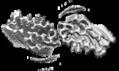 | |||||||||
|---|---|---|---|---|---|---|---|---|---|---|
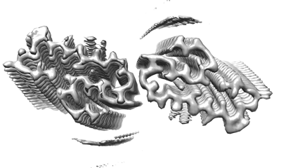
| Title | Cryo-EM structure of the fibril formed by disaccharide-modified amyloid-beta(1-42) | |||||||||
 Map data Map data | ||||||||||
 Sample Sample |
| |||||||||
| Function / homology |  Function and homology information Function and homology informationGolgi-associated vesicle /  clathrin-coated pit / heparin binding / growth cone / perikaryon / early endosome / Golgi apparatus / cell surface / endoplasmic reticulum / extracellular region / nucleus clathrin-coated pit / heparin binding / growth cone / perikaryon / early endosome / Golgi apparatus / cell surface / endoplasmic reticulum / extracellular region / nucleusSimilarity search - Function | |||||||||
| Biological species |  Homo sapiens (human) Homo sapiens (human) | |||||||||
| Method | helical reconstruction / cryo EM / Resolution: 3.1 Å | |||||||||
 Authors Authors | Xia WC / Sun YP / Liu C | |||||||||
| Funding support | 1 items
| |||||||||
 Citation Citation | Journal: J.Am.Chem.Soc. / Year: 2021 Title: O-Glycosylation Induces Amyloid-beta To Form New Fibril Polymorphs Vulnerable for Degradation Authors: Liu D / Wei Q / Xia W / He C / Zhang Q / Huang L / Wang X / Sun Y / Ma Y / Zhang X / Wang Y / Shi X / Liu C / Dong S | |||||||||
| History |
|
- Structure visualization
| Supplemental images |
|---|
- Downloads & links
-EMDB archive
| Map data | emd_31428.map.gz | 4.4 MB | EMDB map data format | |
|---|---|---|---|---|
| Header (meta data) | emd-31428-v30.xmlemd-31428.xml | 9.4 KB 9.4 KB | Display Display | EMDB header |
| FSC (resolution estimation) | emd_31428_fsc.xml | 7.4 KB | Display | FSC data file |
| Images |  emd_31428.png emd_31428.png | 80.4 KB | ||
| Archive directory |  http://ftp.pdbj.org/pub/emdb/structures/EMD-31428ftp://ftp.pdbj.org/pub/emdb/structures/EMD-31428 http://ftp.pdbj.org/pub/emdb/structures/EMD-31428ftp://ftp.pdbj.org/pub/emdb/structures/EMD-31428 | HTTPS FTP |
-Related structure data
| Related structure data | 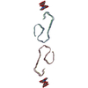 7f29MC M: atomic model generated by this map C: citing same article ( |
|---|---|
| Similar structure data |
-Links
| EMDB pages | EMDB (EBI/PDBe) / EMDataResource |
|---|---|
| Related items in Molecule of the Month |


-Map
| File | Download / File: emd_31428.map.gz / Format: CCP4 / Size: 34.3 MB / Type: IMAGE STORED AS FLOATING POINT NUMBER (4 BYTES) | ||||||||||||||||||||
|---|---|---|---|---|---|---|---|---|---|---|---|---|---|---|---|---|---|---|---|---|---|
| Voxel size | X=Y=Z: 1.06 Å | ||||||||||||||||||||
| Density |
| ||||||||||||||||||||
| Symmetry | Space group: 1 | ||||||||||||||||||||
| Details | EMDB XML:
|
-Supplemental data
- Sample components
-Entire : Cryo-EM structure of the fibril formed by disaccharide-modified a...
| Entire | Name: Cryo-EM structure of the fibril formed by disaccharide-modified amyloid-beta(1-42) |
|---|---|
| Components |
|
-Supramolecule #1: Cryo-EM structure of the fibril formed by disaccharide-modified a...
| Supramolecule | Name: Cryo-EM structure of the fibril formed by disaccharide-modified amyloid-beta(1-42) type: organelle_or_cellular_component / ID: 1 / Parent: 0 / Macromolecule list: #1 |
|---|---|
| Source (natural) | Organism: Homo sapiens (human) |
-Macromolecule #1: Amyloid-beta A4 protein
| Macromolecule | Name: Amyloid-beta A4 protein / type: protein_or_peptide / ID: 1 / Number of copies: 6 / Enantiomer: LEVO |
|---|---|
| Source (natural) | Organism: Homo sapiens (human) |
| Molecular weight | Theoretical: 3.90044 KDa |
| Sequence | String: HDSGYEVHHQ KLVFFAEDVG SNKGAIIGLM VGGVVIA |
-Macromolecule #3: ACETIC ACID
| Macromolecule | Name: ACETIC ACID / type: ligand / ID: 3 / Number of copies: 6 / Formula: ACY |
|---|---|
| Molecular weight | Theoretical: 60.052 Da |
| Chemical component information |  ChemComp-ACY: |
-Experimental details
-Structure determination
| Method | cryo EM |
|---|---|
 Processing Processing | helical reconstruction |
| Aggregation state | filament |
-Sample preparation
| Buffer | pH: 4 |
|---|---|
| Vitrification | Cryogen name: ETHANE |
- Electron microscopy
| Microscope | FEI TITAN KRIOS |
|---|---|
| Electron beam | Acceleration voltage: 300 kV / Electron source: FIELD EMISSION GUN |
| Electron optics | Illumination mode: FLOOD BEAM / Imaging mode: BRIGHT FIELDBright-field microscopy / Nominal defocus max: 2.0 µm / Nominal defocus min: 1.0 µm |
| Image recording | Film or detector model: GATAN K3 (6k x 4k) / Average electron dose: 55.0 e/Å2 |
| Experimental equipment |  Model: Titan Krios / Image courtesy: FEI Company |
-Image processing
| Final angle assignment | Type: NOT APPLICABLE |
|---|---|
| Final reconstruction | Applied symmetry - Helical parameters - Δz: 2.4 Å Applied symmetry - Helical parameters - Δ&Phi: 179.324 ° Applied symmetry - Helical parameters - Axial symmetry: C1 (asymmetric) Resolution.type: BY AUTHOR / Resolution: 3.1 Å / Resolution method: FSC 0.143 CUT-OFF / Number images used: 92523 |
| FSC plot (resolution estimation) | 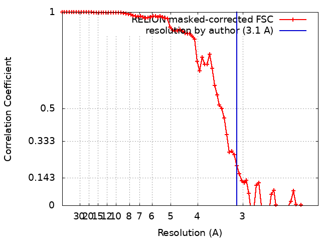 |
