 Movie
Movie Controller

[English] 日本語
 Yorodumi
- EMDB-31325: cryoEM structure of particulate methane monooxygenase associated ... -
+ Open data

- Basic information
| Entry | Database: EMDB / ID: EMD-31325 | |||||||||
|---|---|---|---|---|---|---|---|---|---|---|
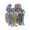
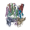
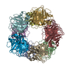
| Title | cryoEM structure of particulate methane monooxygenase associated with Cu(I) | |||||||||
 Map data Map data | ||||||||||
 Sample Sample |
| |||||||||
| Function / homology |  Function and homology information Function and homology information methane monooxygenase (particulate) / methane monooxygenase complex / methane monooxygenase activity / methane monooxygenase (soluble) / methane monooxygenase NADH activity / methane monooxygenase NADPH activity / methane metabolic process / monooxygenase activity / membrane / metal ion binding methane monooxygenase (particulate) / methane monooxygenase complex / methane monooxygenase activity / methane monooxygenase (soluble) / methane monooxygenase NADH activity / methane monooxygenase NADPH activity / methane metabolic process / monooxygenase activity / membrane / metal ion bindingSimilarity search - Function | |||||||||
| Biological species |  Methylococcus capsulatus str. Bath (bacteria) / Methylococcus capsulatus (strain ATCC 33009 / NCIMB 11132 / Bath) (bacteria) Methylococcus capsulatus str. Bath (bacteria) / Methylococcus capsulatus (strain ATCC 33009 / NCIMB 11132 / Bath) (bacteria) | |||||||||
| Method | single particle reconstruction / cryo EM / Resolution: 2.6 Å | |||||||||
 Authors Authors | Chang WH / Lin HH / Tsai IK / Huang SH / Chung SC / Tu IP / Yu SF / Chan SI | |||||||||
 Citation Citation | Journal: J Am Chem Soc / Year: 2021 Title: Copper Centers in the Cryo-EM Structure of Particulate Methane Monooxygenase Reveal the Catalytic Machinery of Methane Oxidation. Authors: W-H Chang / H-H Lin / I-K Tsai / S-H Huang / S-C Chung / I-P Tu / S S-F Yu / S I Chan /  Abstract: The particulate methane monooxygenase (pMMO) is the first enzyme in the C1 metabolic pathway in methanotrophic bacteria. As this enzyme converts methane into methanol efficiently near room ...The particulate methane monooxygenase (pMMO) is the first enzyme in the C1 metabolic pathway in methanotrophic bacteria. As this enzyme converts methane into methanol efficiently near room temperature, it has become the paradigm for developing an understanding of this difficult C1 chemistry. pMMO is a membrane-bound protein with three subunits (PmoB, PmoA, and PmoC) and 12-14 coppers distributed among different sites. X-ray crystal structures that have revealed only three mononuclear coppers at three sites have neither disclosed the location of the active site nor the catalytic mechanism of the enzyme. Here we report a cyro-EM structure of -pMMO from (Bath) at 2.5 Å, and develop quantitative electrostatic-potential profiling to scrutinize the nonprotein densities for signatures of the copper cofactors. Our results confirm a mononuclear Cu at the site, resolve two Cus at the site, and uncover additional Cu clusters at the PmoA/PmoC interface within the membrane ( site) and in the water-exposed -terminal subdomain of the PmoB ( clusters). These findings complete the minimal set of copper factors required for catalytic turnover of pMMO, offering a glimpse of the catalytic machinery for methane oxidation according to the chemical principles underlying the mechanism proposed earlier. | |||||||||
| History |
|
- Structure visualization
| Movie |
Movie viewer |
|---|---|
| Structure viewer | EM map: SurfViewMolmilJmol/JSmol |
| Supplemental images |

- Downloads & links
-EMDB archive
| Map data | emd_31325.map.gz | 306.7 MB | EMDB map data format | |
|---|---|---|---|---|
| Header (meta data) | emd-31325-v30.xmlemd-31325.xml | 18.6 KB 18.6 KB | Display Display | EMDB header |
| FSC (resolution estimation) | emd_31325_fsc.xml | 15.6 KB | Display | FSC data file |
| Images | 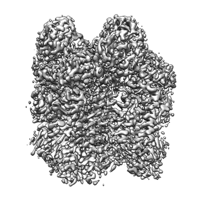 emd_31325.png emd_31325.png | 81.8 KB | ||
| Others | emd_31325_half_map_1.map.gzemd_31325_half_map_2.map.gz | 301.9 MB 301.9 MB | ||
| Archive directory |  http://ftp.pdbj.org/pub/emdb/structures/EMD-31325ftp://ftp.pdbj.org/pub/emdb/structures/EMD-31325 http://ftp.pdbj.org/pub/emdb/structures/EMD-31325ftp://ftp.pdbj.org/pub/emdb/structures/EMD-31325 | HTTPS FTP |
-Related structure data
| Related structure data | 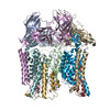 7ev9MC M: atomic model generated by this map C: citing same article ( |
|---|---|
| Similar structure data |


-Links
| EMDB pages | EMDB (EBI/PDBe) / EMDataResource |
|---|
-Map
| File | Download / File: emd_31325.map.gz / Format: CCP4 / Size: 325 MB / Type: IMAGE STORED AS FLOATING POINT NUMBER (4 BYTES) | ||||||||||||||||||||||||||||||||||||||||||||||||||||||||||||
|---|---|---|---|---|---|---|---|---|---|---|---|---|---|---|---|---|---|---|---|---|---|---|---|---|---|---|---|---|---|---|---|---|---|---|---|---|---|---|---|---|---|---|---|---|---|---|---|---|---|---|---|---|---|---|---|---|---|---|---|---|---|
| Voxel size | X=Y=Z: 0.822 Å | ||||||||||||||||||||||||||||||||||||||||||||||||||||||||||||
| Density |
| ||||||||||||||||||||||||||||||||||||||||||||||||||||||||||||
| Symmetry | Space group: 1 | ||||||||||||||||||||||||||||||||||||||||||||||||||||||||||||
| Details | EMDB XML:
CCP4 map header:
| ||||||||||||||||||||||||||||||||||||||||||||||||||||||||||||
-Supplemental data
-Half map: #2
| File | emd_31325_half_map_1.map | ||||||||||||
|---|---|---|---|---|---|---|---|---|---|---|---|---|---|
| Projections & Slices |
| ||||||||||||
| Density Histograms |
 Z
Z Y
Y X
X


-Half map: #1
| File | emd_31325_half_map_2.map | ||||||||||||
|---|---|---|---|---|---|---|---|---|---|---|---|---|---|
| Projections & Slices |
| ||||||||||||
| Density Histograms |


- Sample components
-Entire : particulate methane monooxygenase (pMMO)
| Entire | Name: particulate methane monooxygenase (pMMO) |
|---|---|
| Components |
|
-Supramolecule #1: particulate methane monooxygenase (pMMO)
| Supramolecule | Name: particulate methane monooxygenase (pMMO) / type: complex / ID: 1 / Parent: 0 / Macromolecule list: #1-#3 |
|---|---|
| Source (natural) | Organism: Methylococcus capsulatus str. Bath (bacteria) / Location in cell: membrane |
| Molecular weight | Theoretical: 312 KDa |
-Macromolecule #1: Particulate methane monooxygenase alpha subunit
| Macromolecule | Name: Particulate methane monooxygenase alpha subunit / type: protein_or_peptide / ID: 1 / Number of copies: 3 / Enantiomer: LEVO / EC number: methane monooxygenase (particulate) |
|---|---|
| Source (natural) | Organism: Methylococcus capsulatus (strain ATCC 33009 / NCIMB 11132 / Bath) (bacteria) Strain: ATCC 33009 / NCIMB 11132 / Bath |
| Molecular weight | Theoretical: 46.129746 KDa |
| Sequence | String: MKTIKDRIAK WSAIGLLSAV AATAFYAPSA SAHGEKSQAA FMRMRTIHWY DLSWSKEKVK INETVEIKGK FHVFEGWPET VDEPDVAFL NVGMPGPVFI RKESYIGGQL VPRSVRLEIG KTYDFRVVLK ARRPGDWHVH TMMNVQGGGP IIGPGKWITV E GSMSEFRN ...String: MKTIKDRIAK WSAIGLLSAV AATAFYAPSA SAHGEKSQAA FMRMRTIHWY DLSWSKEKVK INETVEIKGK FHVFEGWPET VDEPDVAFL NVGMPGPVFI RKESYIGGQL VPRSVRLEIG KTYDFRVVLK ARRPGDWHVH TMMNVQGGGP IIGPGKWITV E GSMSEFRN PVTTLTGQTV DLENYNEGNT YFWHAFWFAI GVAWIGYWSR RPIFIPRLLM VDAGRADELV SATDRKVAMG FL AATILIV VMAMSSANSK YPITIPLQAG TMRGMKPLEL PAPTVSVKVE DATYRVPGRA MRMKLTITNH GNSPIRLGEF YTA SVRFLD SDVYKDTTGY PEDLLAEDGL SVSDNSPLAP GETRTVDVTA SDAAWEVYRL SDIIYDPDSR FAGLLFFFDA TGNR QVVQI DAPLIPSFM |
-Macromolecule #2: Particulate methane monooxygenase beta subunit
| Macromolecule | Name: Particulate methane monooxygenase beta subunit / type: protein_or_peptide / ID: 2 / Number of copies: 3 / Enantiomer: LEVO / EC number: methane monooxygenase (particulate) |
|---|---|
| Source (natural) | Organism: Methylococcus capsulatus (strain ATCC 33009 / NCIMB 11132 / Bath) (bacteria) Strain: ATCC 33009 / NCIMB 11132 / Bath |
| Molecular weight | Theoretical: 28.445098 KDa |
| Sequence | String: MSAAQSAVRS HAEAVQVSRT IDWMALFVVF FVIVGSYHIH AMLTMGDWDF WSDWKDRRLW VTVTPIVLVT FPAAVQSYLW ERYRLPWGA TVCVLGLLLG EWINRYFNFW GWTYFPINFV FPASLVPGAI ILDTVLMLSG SYLFTAIVGA MGWGLIFYPG N WPIIAPLH ...String: MSAAQSAVRS HAEAVQVSRT IDWMALFVVF FVIVGSYHIH AMLTMGDWDF WSDWKDRRLW VTVTPIVLVT FPAAVQSYLW ERYRLPWGA TVCVLGLLLG EWINRYFNFW GWTYFPINFV FPASLVPGAI ILDTVLMLSG SYLFTAIVGA MGWGLIFYPG N WPIIAPLH VPVEYNGMLM SIADIQGYNY VRTGTPEYIR MVEKGTLRTF GKDVAPVSAF FSAFMSILIY FMWHFIGRWF SN ERFLQST |
-Macromolecule #3: Ammonia monooxygenase/methane monooxygenase, subunit C family protein
| Macromolecule | Name: Ammonia monooxygenase/methane monooxygenase, subunit C family protein type: protein_or_peptide / ID: 3 / Number of copies: 3 / Enantiomer: LEVO / EC number: methane monooxygenase (soluble) |
|---|---|
| Source (natural) | Organism: Methylococcus capsulatus (strain ATCC 33009 / NCIMB 11132 / Bath) (bacteria) Strain: ATCC 33009 / NCIMB 11132 / Bath |
| Molecular weight | Theoretical: 29.839309 KDa |
| Sequence | String: MAATTIGGAA AAEAPLLDKK WLTFALAIYT VFYLWVRWYE GVYGWSAGLD SFAPEFETYW MNFLYTEIVL EIVTASILWG YLWKTRDRN LAALTPREEL RRNFTHLVWL VAYAWAIYWG ASYFTEQDGT WHQTIVRDTD FTPSHIIEFY LSYPIYIITG F AAFIYAKT ...String: MAATTIGGAA AAEAPLLDKK WLTFALAIYT VFYLWVRWYE GVYGWSAGLD SFAPEFETYW MNFLYTEIVL EIVTASILWG YLWKTRDRN LAALTPREEL RRNFTHLVWL VAYAWAIYWG ASYFTEQDGT WHQTIVRDTD FTPSHIIEFY LSYPIYIITG F AAFIYAKT RLPFFAKGIS LPYLVLVVGP FMILPNVGLN EWGHTFWFME ELFVAPLHYG FVIFGWLALA VMGTLTQTFY SF AQGGLGQ SLCEAVDEGL IAK |
-Macromolecule #4: COPPER (I) ION
| Macromolecule | Name: COPPER (I) ION / type: ligand / ID: 4 / Number of copies: 24 / Formula: CU1 |
|---|---|
| Molecular weight | Theoretical: 63.546 Da |
| Chemical component information |  ChemComp-CU1: |
-Experimental details
-Structure determination
| Method | cryo EM |
|---|---|
 Processing Processing | single particle reconstruction |
| Aggregation state | particle |
-Sample preparation
| Buffer | pH: 7.2 |
|---|---|
| Sugar embedding | Material: vitrified ice |
| Vitrification | Cryogen name: ETHANE / Chamber humidity: 98 % / Chamber temperature: 278 K |
- Electron microscopy
| Microscope | FEI TITAN KRIOS |
|---|---|
| Electron beam | Acceleration voltage: 300 kV / Electron source: FIELD EMISSION GUN |
| Electron optics | Illumination mode: FLOOD BEAM / Imaging mode: BRIGHT FIELDBright-field microscopy / Cs: 2.7 mm / Nominal defocus max: 2.2 µm / Nominal defocus min: 1.0 µm / Nominal magnification: 165000 |
| Sample stage | Cooling holder cryogen: NITROGEN |
| Image recording | Film or detector model: GATAN K2 SUMMIT (4k x 4k) / Detector mode: COUNTING / Digitization - Frames/image: 1-60 / Number real images: 23484 / Average electron dose: 80.0 e/Å2 |
| Experimental equipment |  Model: Titan Krios / Image courtesy: FEI Company |
-Image processing
| CTF correction | Software - Name: Gctf (ver. 1.18) |
|---|---|
| Startup model | Type of model: OTHER / Details: cryoSPARC ab-initio Reconstruction |
| Initial angle assignment | Type: RANDOM ASSIGNMENT / Software - Name: cryoSPARC (ver. 2.15) |
| Final angle assignment | Type: MAXIMUM LIKELIHOOD / Software: (Name: RELION (ver. 3.0), cryoSPARC (ver. 2.15)) |
| Final reconstruction | Applied symmetry - Point group: C3 (3 fold cyclic) / Resolution.type: BY AUTHOR / Resolution: 2.6 Å / Resolution method: FSC 0.143 CUT-OFF / Software - Name: cryoSPARC (ver. 2.15) / Number images used: 128116 |
| FSC plot (resolution estimation) |  |
