Oxidoreductases; Acting on the CH-CH group of donors / tricarboxylic acid cycle / 2 iron, 2 sulfur cluster binding / 4 iron, 4 sulfur cluster binding / membrane => GO:0016020 / electron transfer activity / oxidoreductase activity / metal ion binding Similarity search - Function
National Natural Science Foundation of China (NSFC)
81520108019, 813300237
China
Chinese Academy of Sciences
2017YFC0840300
China
Citation
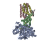
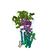
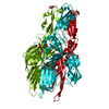
Journal: Proc Natl Acad Sci U S A / Year: 2021 Title: Architecture of the mycobacterial succinate dehydrogenase with a membrane-embedded Rieske FeS cluster. Authors: Xiaoting Zhou / Yan Gao / Weiwei Wang / Xiaolin Yang / Xiuna Yang / Fengjiang Liu / Yanting Tang / Sin Man Lam / Guanghou Shui / Lu Yu / Changlin Tian / Luke W Guddat / Quan Wang / Zihe Rao / Hongri Gong / Abstract: Complex II, also known as succinate dehydrogenase (SQR) or fumarate reductase (QFR), is an enzyme involved in both the Krebs cycle and oxidative phosphorylation. Mycobacterial Sdh1 has recently been ...Complex II, also known as succinate dehydrogenase (SQR) or fumarate reductase (QFR), is an enzyme involved in both the Krebs cycle and oxidative phosphorylation. Mycobacterial Sdh1 has recently been identified as a new class of respiratory complex II (type F) but with an unknown electron transfer mechanism. Here, using cryoelectron microscopy, we have determined the structure of Sdh1 in the presence and absence of the substrate, ubiquinone-1, at 2.53-Å and 2.88-Å resolution, respectively. Sdh1 comprises three subunits, two that are water soluble, SdhA and SdhB, and one that is membrane spanning, SdhC. Within these subunits we identified a quinone-binding site and a rarely observed Rieske-type [2Fe-2S] cluster, the latter being embedded in the transmembrane region. A mutant, where two His ligands of the Rieske-type [2Fe-2S] were changed to alanine, abolished the quinone reduction activity of the Sdh1. Our structures allow the proposal of an electron transfer pathway that connects the substrate-binding and quinone-binding sites. Given the unique features of Sdh1 and its essential role in , these structures will facilitate antituberculosis drug discovery efforts that specifically target this complex.
History
Deposition
Oct 2, 2020
-
Header (metadata) release
Apr 7, 2021
-
Map release
Apr 7, 2021
-
Update
Feb 16, 2022
-
Current status
Feb 16, 2022
Processing site: PDBj / Status: Released
-
Structure visualization
Movie
Surface view with section colored by density value
Macromolecule #1: Succinate dehydrogenase subunit A
Macromolecule
Name: Succinate dehydrogenase subunit A / type: protein_or_peptide / ID: 1 / Number of copies: 1 / Enantiomer: LEVO EC number: Oxidoreductases; Acting on the CH-CH group of donors
In the structure databanks used in Yorodumi, some data are registered as the other names, "COVID-19 virus" and "2019-nCoV". Here are the details of the virus and the list of structure data.
Jan 31, 2019. EMDB accession codes are about to change! (news from PDBe EMDB page)
EMDB accession codes are about to change! (news from PDBe EMDB page)
The allocation of 4 digits for EMDB accession codes will soon come to an end. Whilst these codes will remain in use, new EMDB accession codes will include an additional digit and will expand incrementally as the available range of codes is exhausted. The current 4-digit format prefixed with “EMD-” (i.e. EMD-XXXX) will advance to a 5-digit format (i.e. EMD-XXXXX), and so on. It is currently estimated that the 4-digit codes will be depleted around Spring 2019, at which point the 5-digit format will come into force.
The EM Navigator/Yorodumi systems omit the EMD- prefix.
Related info.:Q: What is EMD? / ID/Accession-code notation in Yorodumi/EM Navigator
Yorodumi is a browser for structure data from EMDB, PDB, SASBDB, etc.
This page is also the successor to EM Navigator detail page, and also detail information page/front-end page for Omokage search.
The word "yorodu" (or yorozu) is an old Japanese word meaning "ten thousand". "mi" (miru) is to see.
Related info.:EMDB / PDB / SASBDB / Comparison of 3 databanks / Yorodumi Search / Aug 31, 2016. New EM Navigator & Yorodumi / Yorodumi Papers / Jmol/JSmol / Function and homology information / Changes in new EM Navigator and Yorodumi
 Movie
Movie Controller


 Open data
 Basic information
 Map data
 Sample
 Function and homology information
 Oxidoreductases; Acting on the CH-CH group of donors /
  Mycolicibacterium smegmatis MC2 51 (bacteria) /
  Authors
 China, 3 items
  Citation
 Structure visualization
 Downloads & links
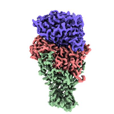 emd_30594.png
 http://ftp.pdbj.org/pub/emdb/structures/EMD-30594


 Sample components


 Processing
 Electron microscopy
