 Movie
Movie Controller

[English] 日本語
 Yorodumi
- EMDB-29780: Cryo-Electron Tomography of a Cryo-Lamella cut inside a Mouse Kid... -
+ Open data

- Basic information
| Entry |  | ||||||||||||
|---|---|---|---|---|---|---|---|---|---|---|---|---|---|
| Title | Cryo-Electron Tomography of a Cryo-Lamella cut inside a Mouse Kidney Tissue Biopsy | ||||||||||||
 Map data Map data | Cryo-Electron Tomography of Mouse Kidney Tissue Biopsy | ||||||||||||
 Sample Sample |
| ||||||||||||
 Keywords Keywords |  Microtubules / mitochondria / UNKNOWN FUNCTION Microtubules / mitochondria / UNKNOWN FUNCTION | ||||||||||||
| Biological species |  Mus musculus (house mouse) Mus musculus (house mouse) | ||||||||||||
| Method | electron tomography | ||||||||||||
 Authors Authors | Trepout S / Ramm G | ||||||||||||
| Funding support |  Australia, Australia,  United States, 3 items United States, 3 items
| ||||||||||||
 Citation Citation | Journal: To Be Published Title: Cryo-Electron Tomography of a Cryo-Lamella Milled Inside a Mouse Kidney Tissue Biopsy Authors: Trepout S / Ramm G | ||||||||||||
| History |
|
- Structure visualization
| Supplemental images |
|---|
- Downloads & links
-EMDB archive
| Map data | emd_29780.map.gz | 4.3 GB |  EMDB map data format EMDB map data format | |
|---|---|---|---|---|
| Header (meta data) | emd-29780-v30.xmlemd-29780.xml | 8.9 KB 8.9 KB | Display Display | EMDB header |
| Images |  emd_29780.png emd_29780.png | 236.5 KB | ||
| Filedesc metadata | emd-29780.cif.gz | 3.7 KB | ||
| Archive directory |  http://ftp.pdbj.org/pub/emdb/structures/EMD-29780ftp://ftp.pdbj.org/pub/emdb/structures/EMD-29780 http://ftp.pdbj.org/pub/emdb/structures/EMD-29780ftp://ftp.pdbj.org/pub/emdb/structures/EMD-29780 | HTTPS FTP |
-Links
| EMDB pages | EMDB (EBI/PDBe) / EMDataResource |
|---|
-Map
| File | Download / File: emd_29780.map.gz / Format: CCP4 / Size: 4.6 GB / Type: IMAGE STORED AS FLOATING POINT NUMBER (4 BYTES) | ||||||||||||||||||||||||||||||||
|---|---|---|---|---|---|---|---|---|---|---|---|---|---|---|---|---|---|---|---|---|---|---|---|---|---|---|---|---|---|---|---|---|---|
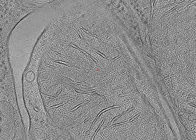
| Annotation | Cryo-Electron Tomography of Mouse Kidney Tissue Biopsy | ||||||||||||||||||||||||||||||||
| Projections & slices | Image control
Images are generated by Spider. generated in cubic-lattice coordinate | ||||||||||||||||||||||||||||||||
| Voxel size | X=Y=Z: 14.66 Å | ||||||||||||||||||||||||||||||||
| Density |
| ||||||||||||||||||||||||||||||||
| Symmetry | Space group: 1 | ||||||||||||||||||||||||||||||||
| Details | EMDB XML:
|
 Z (Sec.)
 Y (Row.)
 X (Col.)


-Supplemental data
- Sample components
-Entire : Biopsy of Mouse Kidney Tissue
| Entire | Name: Biopsy of Mouse Kidney Tissue |
|---|---|
| Components |
|
-Supramolecule #1: Biopsy of Mouse Kidney Tissue
| Supramolecule | Name: Biopsy of Mouse Kidney Tissue / type: tissue / ID: 1 / Parent: 0 Details: Cryo-lamella cut inside the biopsy using the bbq method. |
|---|---|
| Source (natural) | Organism: Mus musculus (house mouse) / Organ: kidney |
-Experimental details
-Structure determination
 Processing Processing | electron tomography |
|---|---|
| Aggregation state | tissue |
-Sample preparation
| Buffer | pH: 6.9 |
|---|---|
| High pressure freezing | Instrument: OTHER Details: The value given for _em_high_pressure_freezing.instrument is HPF Wohlwend Compact 03. This is not in a list of allowed values {'LEICA EM HPM100', 'LEICA EM PACT2', 'OTHER', 'EMS-002 RAPID ...Details: The value given for _em_high_pressure_freezing.instrument is HPF Wohlwend Compact 03. This is not in a list of allowed values {'LEICA EM HPM100', 'LEICA EM PACT2', 'OTHER', 'EMS-002 RAPID IMMERSION FREEZER', 'LEICA EM PACT', 'BAL-TEC HPM 010'} so OTHER is written into the XML file. |
| Sectioning | Focused ion beam - Instrument: OTHER / Focused ion beam - Ion: OTHER / Focused ion beam - Voltage: 30 / Focused ion beam - Current: 1 / Focused ion beam - Duration: 1800 / Focused ion beam - Temperature: 100 K / Focused ion beam - Initial thickness: 100000 / Focused ion beam - Final thickness: 250 Focused ion beam - Details: The value given for _em_focused_ion_beam.instrument is FEI Helios G4 UX. This is not in a list of allowed values {'OTHER', 'DB235'} so OTHER is written into the XML file. |
- Electron microscopy
| Microscope | FEI TITAN KRIOS |
|---|---|
| Electron beam | Acceleration voltage: 300 kV / Electron source: FIELD EMISSION GUN |
| Electron optics | Illumination mode: FLOOD BEAM / Imaging mode: BRIGHT FIELDBright-field microscopy / Nominal defocus max: 4.0 µm / Nominal defocus min: 4.0 µm |
| Specialist optics | Energy filter - Name: GIF Bioquantum / Energy filter - Slit width: 20 eV |
| Sample stage | Specimen holder model: FEI TITAN KRIOS AUTOGRID HOLDER / Cooling holder cryogen: NITROGEN |
| Image recording | Film or detector model: GATAN K2 QUANTUM (4k x 4k) / Detector mode: COUNTING / Average electron dose: 1.5 e/Å2 |
| Experimental equipment |  Model: Titan Krios / Image courtesy: FEI Company |
-Image processing
| Final reconstruction | Algorithm: BACK PROJECTION / Software - Name: IMOD / Details: Tilt-series alignment performed in AreTomo v1.0.1 / Number images used: 59 |
|---|
