National Institutes of Health/National Institute of General Medical Sciences (NIH/NIGMS)
R01GM143380
United States
National Institutes of Health/National Heart, Lung, and Blood Institute (NIH/NHLBI)
R01HL162842
United States
National Institutes of Health/National Institute of General Medical Sciences (NIH/NIGMS)
R01GM080139
United States
Cancer Prevention and Research Institute of Texas (CPRIT)
RP190602
United States
Citation
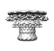
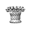
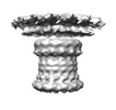
Journal: Nat Commun / Year: 2023 Title: Membrane translocation process revealed by in situ structures of type II secretion system secretins. Authors: Zhili Yu / Yaoming Wu / Muyuan Chen / Tong Huo / Wei Zheng / Steven J Ludtke / Xiaodong Shi / Zhao Wang / Abstract: The GspD secretin is the outer membrane channel of the bacterial type II secretion system (T2SS) which secrets diverse toxins that cause severe diseases such as diarrhea and cholera. GspD needs to ...The GspD secretin is the outer membrane channel of the bacterial type II secretion system (T2SS) which secrets diverse toxins that cause severe diseases such as diarrhea and cholera. GspD needs to translocate from the inner to the outer membrane to exert its function, and this process is an essential step for T2SS to assemble. Here, we investigate two types of secretins discovered so far in Escherichia coli, GspD, and GspD. By electron cryotomography subtomogram averaging, we determine in situ structures of key intermediate states of GspD and GspD in the translocation process, with resolution ranging from 9 Å to 19 Å. In our results, GspD and GspD present entirely different membrane interaction patterns and ways of transitioning the peptidoglycan layer. From this, we hypothesize two distinct models for the membrane translocation of GspD and GspD, providing a comprehensive perspective on the inner to outer membrane biogenesis of T2SS secretins.
In the structure databanks used in Yorodumi, some data are registered as the other names, "COVID-19 virus" and "2019-nCoV". Here are the details of the virus and the list of structure data.
Jan 31, 2019. EMDB accession codes are about to change! (news from PDBe EMDB page)
EMDB accession codes are about to change! (news from PDBe EMDB page)
The allocation of 4 digits for EMDB accession codes will soon come to an end. Whilst these codes will remain in use, new EMDB accession codes will include an additional digit and will expand incrementally as the available range of codes is exhausted. The current 4-digit format prefixed with “EMD-” (i.e. EMD-XXXX) will advance to a 5-digit format (i.e. EMD-XXXXX), and so on. It is currently estimated that the 4-digit codes will be depleted around Spring 2019, at which point the 5-digit format will come into force.
The EM Navigator/Yorodumi systems omit the EMD- prefix.
Related info.:Q: What is EMD? / ID/Accession-code notation in Yorodumi/EM Navigator
Yorodumi is a browser for structure data from EMDB, PDB, SASBDB, etc.
This page is also the successor to EM Navigator detail page, and also detail information page/front-end page for Omokage search.
The word "yorodu" (or yorozu) is an old Japanese word meaning "ten thousand". "mi" (miru) is to see.
Related info.:EMDB / PDB / SASBDB / Comparison of 3 databanks / Yorodumi Search / Aug 31, 2016. New EM Navigator & Yorodumi / Yorodumi Papers / Jmol/JSmol / Function and homology information / Changes in new EM Navigator and Yorodumi
 Movie
Movie Controller

 Yorodumi
 Open data
 Basic information
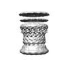
 Map data
 Sample
 Keywords
 MEMBRANE PROTEIN
 Authors
 United States, 4 items
  Citation
 Structure visualization
 Downloads & links
 EMDB map data format
 emd_29702.png
 http://ftp.pdbj.org/pub/emdb/structures/EMD-29702
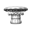
 Z
Z Y
Y X
X


 Sample components
 Processing
 Electron microscopy

