 Movie
Movie Controller

[English] 日本語
 Yorodumi
- EMDB-29485: E. coli 70S ribosome with an improved MS2 tag inserted in H98 (30... -
+ Open data

- Basic information
| Entry | 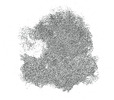 | |||||||||
|---|---|---|---|---|---|---|---|---|---|---|
| Title | E. coli 70S ribosome with an improved MS2 tag inserted in H98 (30S focused map) | |||||||||
 Map data Map data | E. coli 70S ribosome with an improved MS2 tag inserted in H98 (30S focused map) | |||||||||
 Sample Sample |
| |||||||||
 Keywords Keywords | MS2-tag / H98 /  ribosome ribosome | |||||||||
| Biological species |  Escherichia coli (E. coli) Escherichia coli (E. coli) | |||||||||
| Method | single particle reconstruction / cryo EM / Resolution: 1.98 Å | |||||||||
 Authors Authors | Nissley AJ / Cate JHD | |||||||||
| Funding support |  United States, 1 items United States, 1 items
| |||||||||
 Citation Citation | Journal: RNA / Year: 2023 Title: Interactions between terminal ribosomal RNA helices stabilize the large ribosomal subunit. Authors: Amos J Nissley / Tammam S Kamal / Jamie H D Cate / Abstract: The ribosome is a large ribonucleoprotein assembly that uses diverse and complex molecular interactions to maintain proper folding. In vivo assembled ribosomes have been isolated using MS2 tags ...The ribosome is a large ribonucleoprotein assembly that uses diverse and complex molecular interactions to maintain proper folding. In vivo assembled ribosomes have been isolated using MS2 tags installed in either the 16S or 23S ribosomal RNAs (rRNAs), to enable studies of ribosome structure and function in vitro. RNA tags in the 50S subunit have commonly been inserted into an extended helix H98 in 23S rRNA, as this addition does not affect cellular growth or in vitro ribosome activity. Here, we find that 50S subunits with MS2 tags inserted in H98 are destabilized compared to wild-type (WT) 50S subunits. We identify the loss of RNA-RNA tertiary contacts that bridge helices H1, H94, and H98 as the cause of destabilization. Using cryogenic electron microscopy (cryo-EM), we show that this interaction is disrupted by the addition of the MS2 tag and can be restored through the insertion of a single adenosine in the extended H98 helix. This work establishes ways to improve MS2 tags in the 50S subunit that maintain ribosome stability and investigates a complex RNA tertiary structure that may be important for stability in various bacterial ribosomes. | |||||||||
| History |
|
- Structure visualization
| Supplemental images |
|---|

- Downloads & links
-EMDB archive
| Map data | emd_29485.map.gz | 324 MB |  EMDB map data format EMDB map data format | |
|---|---|---|---|---|
| Header (meta data) | emd-29485-v30.xmlemd-29485.xml | 15.6 KB 15.6 KB | Display Display | EMDB header |
| FSC (resolution estimation) | emd_29485_fsc.xml | 14.7 KB | Display | FSC data file |
| Images | 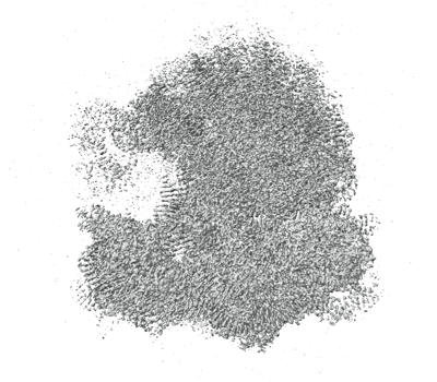 emd_29485.png emd_29485.png | 172.2 KB | ||
| Filedesc metadata | emd-29485.cif.gz | 4.1 KB | ||
| Others | emd_29485_half_map_1.map.gzemd_29485_half_map_2.map.gz | 318 MB 318 MB | ||
| Archive directory |  http://ftp.pdbj.org/pub/emdb/structures/EMD-29485ftp://ftp.pdbj.org/pub/emdb/structures/EMD-29485 http://ftp.pdbj.org/pub/emdb/structures/EMD-29485ftp://ftp.pdbj.org/pub/emdb/structures/EMD-29485 | HTTPS FTP |
-Related structure data

-Links
| EMDB pages | EMDB (EBI/PDBe) / EMDataResource |
|---|
-Map
| File | Download / File: emd_29485.map.gz / Format: CCP4 / Size: 343 MB / Type: IMAGE STORED AS FLOATING POINT NUMBER (4 BYTES) | ||||||||||||||||||||
|---|---|---|---|---|---|---|---|---|---|---|---|---|---|---|---|---|---|---|---|---|---|
| Annotation | E. coli 70S ribosome with an improved MS2 tag inserted in H98 (30S focused map) | ||||||||||||||||||||
| Voxel size | X=Y=Z: 0.8248 Å | ||||||||||||||||||||
| Density |
| ||||||||||||||||||||
| Symmetry | Space group: 1 | ||||||||||||||||||||
| Details | EMDB XML:
|
-Supplemental data
-Half map: E. coli 70S ribosome with an improved MS2...
| File | emd_29485_half_map_1.map | ||||||||||||
|---|---|---|---|---|---|---|---|---|---|---|---|---|---|
| Annotation | E. coli 70S ribosome with an improved MS2 tag inserted in H98 (30S focused half map) | ||||||||||||
| Projections & Slices |
| ||||||||||||
| Density Histograms |
 Z
Z Y
Y X
X


-Half map: E. coli 70S ribosome with an improved MS2...
| File | emd_29485_half_map_2.map | ||||||||||||
|---|---|---|---|---|---|---|---|---|---|---|---|---|---|
| Annotation | E. coli 70S ribosome with an improved MS2 tag inserted in H98 (30S focused half map) | ||||||||||||
| Projections & Slices |
| ||||||||||||
| Density Histograms |


- Sample components
-Entire : E. coli 70S ribosome
| Entire | Name: E. coli 70S ribosome |
|---|---|
| Components |
|
-Supramolecule #1: E. coli 70S ribosome
| Supramolecule | Name: E. coli 70S ribosome / type: complex / ID: 1 / Parent: 0 |
|---|---|
| Source (natural) | Organism: Escherichia coli (E. coli) |
-Supramolecule #2: 50S Subunit
| Supramolecule | Name: 50S Subunit / type: complex / ID: 2 / Parent: 1 |
|---|---|
| Source (natural) | Organism: Escherichia coli (E. coli) |
-Supramolecule #3: 30S Subunit
| Supramolecule | Name: 30S Subunit / type: complex / ID: 3 / Parent: 1 |
|---|---|
| Source (natural) | Organism: Escherichia coli (E. coli) |
-Experimental details
-Structure determination
| Method | cryo EM |
|---|---|
 Processing Processing | single particle reconstruction |
| Aggregation state | particle |
-Sample preparation
| Concentration | 0.27 mg/mL | ||||||||||||
|---|---|---|---|---|---|---|---|---|---|---|---|---|---|
| Buffer | pH: 7.5 Component:
| ||||||||||||
| Grid | Model: UltrAuFoil R1.2/1.3 / Material: GOLD / Mesh: 300 / Support film - Material: CARBON / Support film - topology: CONTINUOUS / Pretreatment - Type: GLOW DISCHARGE | ||||||||||||
| Vitrification | Cryogen name: ETHANE / Chamber humidity: 100 % / Chamber temperature: 277 K / Instrument: FEI VITROBOT MARK III |
- Electron microscopy
| Microscope | FEI TITAN KRIOS |
|---|---|
| Electron beam | Acceleration voltage: 300 kV / Electron source: FIELD EMISSION GUN |
| Electron optics | Illumination mode: OTHER / Imaging mode: BRIGHT FIELDBright-field microscopy / Nominal defocus max: 1.5 µm / Nominal defocus min: 0.5 µm |
| Image recording | Film or detector model: GATAN K3 (6k x 4k) / Number grids imaged: 1 / Number real images: 11736 / Average electron dose: 40.0 e/Å2 |
| Experimental equipment |  Model: Titan Krios / Image courtesy: FEI Company |
-Image processing
| Particle selection | Number selected: 2475999 |
|---|---|
| Startup model | Type of model: OTHER |
| Initial angle assignment | Type: MAXIMUM LIKELIHOOD / Software - Name: cryoSPARC (ver. 3) |
| Final 3D classification | Software - Name: cryoSPARC (ver. 3) |
| Final angle assignment | Type: MAXIMUM LIKELIHOOD / Software - Name: cryoSPARC (ver. 3) |
| Final reconstruction | Resolution.type: BY AUTHOR / Resolution: 1.98 Å / Resolution method: FSC 0.143 CUT-OFF / Software - Name: cryoSPARC (ver. 3) / Number images used: 601617 |
| FSC plot (resolution estimation) |  |
-Atomic model buiding 1
| Refinement | Protocol: RIGID BODY FIT |
|---|
