Movie
Movie Controller

[English] 日本語
 Yorodumi
- EMDB-29380: LBD conformation 1 (LBDconf1) of GluA2 flip Q isoform of AMPA rec... -
+ Open data

- Basic information
| Entry |  | |||||||||
|---|---|---|---|---|---|---|---|---|---|---|
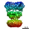
| Title | LBD conformation 1 (LBDconf1) of GluA2 flip Q isoform of AMPA receptor in complex with gain-of-function TARP gamma2, with 140mM NMDG, 330uM CTZ, and 100mM L-glutamate (Open-Na110) | |||||||||
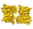
 Map data Map data | LBDconf1, Open-NMDG | |||||||||
 Sample Sample |
| |||||||||
 Keywords Keywords | Inotropic glutamate receptors / AMPA receptors /  ligand gated ion channel / auxiliary subunit / TARP / stargazin / TARP gamma2 / glutamate / calcium / neurotransmitter receptor / synaptic transmission / TRANSPORT PROTEIN ligand gated ion channel / auxiliary subunit / TARP / stargazin / TARP gamma2 / glutamate / calcium / neurotransmitter receptor / synaptic transmission / TRANSPORT PROTEIN | |||||||||
| Function / homology |  Function and homology information Function and homology informationspine synapse / dendritic spine neck / dendritic spine head / Activation of AMPA receptors / response to lithium ion / perisynaptic space / cellular response to glycine / AMPA glutamate receptor activity / Trafficking of GluR2-containing AMPA receptors / immunoglobulin binding ...spine synapse / dendritic spine neck / dendritic spine head / Activation of AMPA receptors / response to lithium ion / perisynaptic space / cellular response to glycine / AMPA glutamate receptor activity / Trafficking of GluR2-containing AMPA receptors / immunoglobulin binding / AMPA glutamate receptor complex / kainate selective glutamate receptor activity / ionotropic glutamate receptor complex / extracellularly glutamate-gated ion channel activity / asymmetric synapse / regulation of receptor recycling / Unblocking of NMDA receptors, glutamate binding and activation / glutamate receptor binding / positive regulation of synaptic transmission / glutamate-gated receptor activity / presynaptic active zone membrane / response to fungicide / regulation of synaptic transmission, glutamatergic / cellular response to brain-derived neurotrophic factor stimulus / somatodendritic compartment / dendrite membrane / ligand-gated monoatomic ion channel activity involved in regulation of presynaptic membrane potential / ionotropic glutamate receptor binding / ionotropic glutamate receptor signaling pathway / dendrite cytoplasm / cytoskeletal protein binding / SNARE binding / transmitter-gated monoatomic ion channel activity involved in regulation of postsynaptic membrane potential / dendritic shaft / synaptic membrane / synaptic transmission, glutamatergic / PDZ domain binding / postsynaptic density membrane / protein tetramerization / modulation of chemical synaptic transmission / Schaffer collateral - CA1 synapse / establishment of protein localization / terminal bouton / receptor internalization / synaptic vesicle membrane / cerebral cortex development / synaptic vesicle / presynapse / presynaptic membrane / signaling receptor activity / amyloid-beta binding / growth cone / perikaryon / chemical synaptic transmission / scaffold protein binding / postsynaptic membrane / dendritic spine / postsynaptic density / neuron projection / axon / neuronal cell body / dendrite / synapse / glutamatergic synapse / protein-containing complex binding / endoplasmic reticulum membrane / protein kinase binding / cell surface / endoplasmic reticulum / protein-containing complex / membrane / identical protein binding / plasma membraneSimilarity search - Function | |||||||||
| Biological species |  Rattus norvegicus (Norway rat) Rattus norvegicus (Norway rat) | |||||||||
| Method | single particle reconstruction / cryo EM / Resolution: 3.11 Å | |||||||||
 Authors Authors | Nakagawa T | |||||||||
| Funding support |  United States, 1 items United States, 1 items
| |||||||||
 Citation Citation | Journal: Nat Struct Mol Biol / Year: 2024 Title: The open gate of the AMPA receptor forms a Ca binding site critical in regulating ion transport. Authors: Terunaga Nakagawa / Xin-Tong Wang / Federico J Miguez-Cabello / Derek Bowie /  Abstract: Alpha-amino-3-hydroxyl-5-methyl-4-isoxazole-propionic acid receptors (AMPARs) are cation-selective ion channels that mediate most fast excitatory neurotransmission in the brain. Although their gating ...Alpha-amino-3-hydroxyl-5-methyl-4-isoxazole-propionic acid receptors (AMPARs) are cation-selective ion channels that mediate most fast excitatory neurotransmission in the brain. Although their gating mechanism has been studied extensively, understanding how cations traverse the pore has remained elusive. Here we investigated putative ion and water densities in the open pore of Ca-permeable AMPARs (rat GRIA2 flip-Q isoform) at 2.3-2.6 Å resolution. We show that the ion permeation pathway attains an extracellular Ca binding site (site-G) when the channel gate moves into the open configuration. Site-G is highly selective for Ca over Na, favoring the movement of Ca into the selectivity filter of the pore. Seizure-related N619K mutation, adjacent to site-G, promotes channel opening but attenuates Ca binding and thus diminishes Ca permeability. Our work identifies the importance of site-G, which coordinates with the Q/R site of the selectivity filter to ensure the preferential transport of Ca through the channel pore. | |||||||||
| History |
|
- Structure visualization
| Supplemental images |
|---|

- Downloads & links
-EMDB archive
| Map data | emd_29380.map.gz | 166.5 MB | EMDB map data format | |
|---|---|---|---|---|
| Header (meta data) | emd-29380-v30.xmlemd-29380.xml | 19.2 KB 19.2 KB | Display Display | EMDB header |
| FSC (resolution estimation) | emd_29380_fsc.xml | 12.8 KB | Display | FSC data file |
| Images | 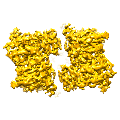 emd_29380.png emd_29380.png | 145.3 KB | ||
| Masks | emd_29380_msk_1.map | 178 MB | Mask map | |
| Filedesc metadata | emd-29380.cif.gz | 6.7 KB | ||
| Others | emd_29380_half_map_1.map.gzemd_29380_half_map_2.map.gz | 139.1 MB 139.1 MB | ||
| Archive directory |  http://ftp.pdbj.org/pub/emdb/structures/EMD-29380ftp://ftp.pdbj.org/pub/emdb/structures/EMD-29380 http://ftp.pdbj.org/pub/emdb/structures/EMD-29380ftp://ftp.pdbj.org/pub/emdb/structures/EMD-29380 | HTTPS FTP |
-Related structure data
| Related structure data | 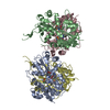 8fq8MC  8fp4C  8fp9C 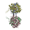 8fpcC  8fpgC 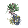 8fphC 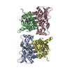 8fpkC 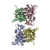 8fplC  8fpsC 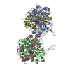 8fpvC 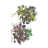 8fpyC 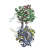 8fpzC  8fq1C 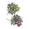 8fq2C 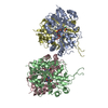 8fq3C  8fq5C 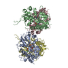 8fq6C 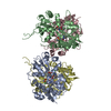 8fqaC  8fqbC 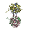 8fqdC 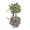 8fqeC  8fqfC 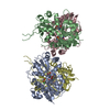 8fqgC 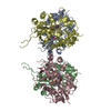 8fqhC  8fr0C C: citing same article ( M: atomic model generated by this map |
|---|---|
| Similar structure data |


-Links
| EMDB pages | EMDB (EBI/PDBe) / EMDataResource |
|---|---|
| Related items in Molecule of the Month |


-Map
| File | Download / File: emd_29380.map.gz / Format: CCP4 / Size: 178 MB / Type: IMAGE STORED AS FLOATING POINT NUMBER (4 BYTES) | ||||||||||||||||||||||||||||||||||||
|---|---|---|---|---|---|---|---|---|---|---|---|---|---|---|---|---|---|---|---|---|---|---|---|---|---|---|---|---|---|---|---|---|---|---|---|---|---|
| Annotation | LBDconf1, Open-NMDG | ||||||||||||||||||||||||||||||||||||
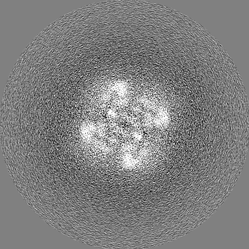
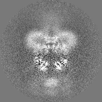
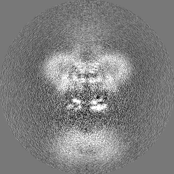
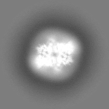
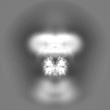
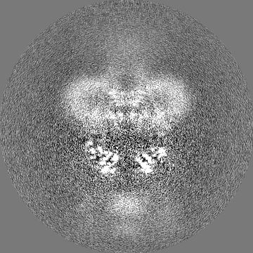
| Projections & slices | Image control
Images are generated by Spider. | ||||||||||||||||||||||||||||||||||||
| Voxel size | X=Y=Z: 0.8195 Å | ||||||||||||||||||||||||||||||||||||
| Density |
| ||||||||||||||||||||||||||||||||||||
| Symmetry | Space group: 1 | ||||||||||||||||||||||||||||||||||||
| Details | EMDB XML:
|
 Z (Sec.)
 Y (Row.)
 X (Col.)


-Supplemental data
-Mask #1
| File | emd_29380_msk_1.map | ||||||||||||
|---|---|---|---|---|---|---|---|---|---|---|---|---|---|
| Projections & Slices |
| ||||||||||||
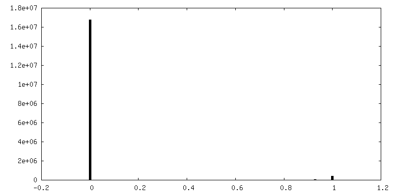
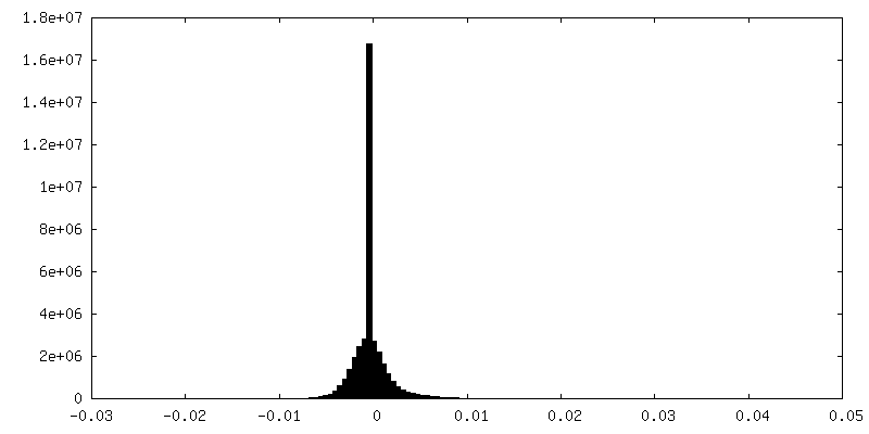
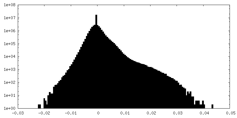
| Density Histograms |


-Half map: Half1 map, Open-NMDG
| File | emd_29380_half_map_1.map | ||||||||||||
|---|---|---|---|---|---|---|---|---|---|---|---|---|---|
| Annotation | Half1 map, Open-NMDG | ||||||||||||
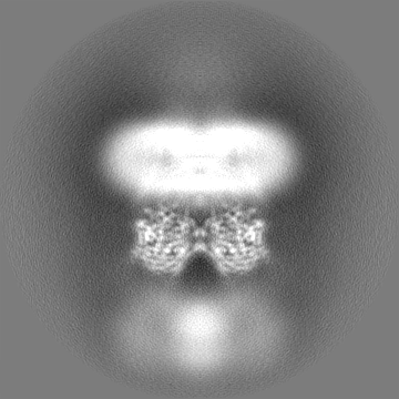
| Projections & Slices |
| ||||||||||||
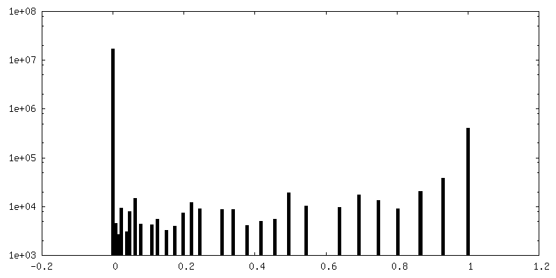
| Density Histograms |

-Half map: Half2 map, Open-NMDG
| File | emd_29380_half_map_2.map | ||||||||||||
|---|---|---|---|---|---|---|---|---|---|---|---|---|---|
| Annotation | Half2 map, Open-NMDG | ||||||||||||
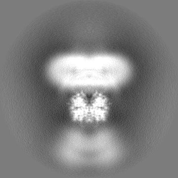
| Projections & Slices |
| ||||||||||||
| Density Histograms |


- Sample components
-Entire : Homotetrameric assembly of GluA2 flip-Q isoform in complex with T...
| Entire | Name: Homotetrameric assembly of GluA2 flip-Q isoform in complex with TARP gamma2 (K52E, K53E). Dimer-of-dimers assembly. |
|---|---|
| Components |
|
-Supramolecule #1: Homotetrameric assembly of GluA2 flip-Q isoform in complex with T...
| Supramolecule | Name: Homotetrameric assembly of GluA2 flip-Q isoform in complex with TARP gamma2 (K52E, K53E). Dimer-of-dimers assembly. type: complex / ID: 1 / Parent: 0 / Macromolecule list: #1 |
|---|---|
| Source (natural) | Organism: Rattus norvegicus (Norway rat) |
| Molecular weight | Theoretical: 500 KDa |
-Macromolecule #1: Glutamate receptor 2
| Macromolecule | Name: Glutamate receptor 2 / type: protein_or_peptide / ID: 1 / Number of copies: 4 / Enantiomer: LEVO |
|---|---|
| Source (natural) | Organism: Rattus norvegicus (Norway rat) |
| Molecular weight | Theoretical: 99.559461 KDa |
| Recombinant expression | Organism:  Homo sapiens (human) Homo sapiens (human) |
| Sequence | String: MQKIMHISVL LSPVLWGLIF GVSSNSIQIG GLFPRGADQE YSAFRVGMVQ FSTSEFRLTP HIDNLEVANS FAVTNAFCSQ FSRGVYAIF GFYDKKSVNT ITSFCGTLHV SFITPSFPTD GTHPFVIQMR PDLKGALLSL IEYYQWDKFA YLYDSDRGLS T LQAVLDSA ...String: MQKIMHISVL LSPVLWGLIF GVSSNSIQIG GLFPRGADQE YSAFRVGMVQ FSTSEFRLTP HIDNLEVANS FAVTNAFCSQ FSRGVYAIF GFYDKKSVNT ITSFCGTLHV SFITPSFPTD GTHPFVIQMR PDLKGALLSL IEYYQWDKFA YLYDSDRGLS T LQAVLDSA AEKKWQVTAI NVGNINNDKK DETYRSLFQD LELKKERRVI LDCERDKVND IVDQVITIGK HVKGYHYIIA NL GFTDGDL LKIQFGGANV SGFQIVDYDD SLVSKFIERW STLEEKEYPG AHTATIKYTS ALTYDAVQVM TEAFRNLRKQ RIE ISRRGN AGDCLANPAV PWGQGVEIER ALKQVQVEGL SGNIKFDQNG KRINYTINIM ELKTNGPRKI GYWSEVDKMV VTLT ELPSG NDTSGLENKT VVVTTILESP YVMMKKNHEM LEGNERYEGY CVDLAAEIAK HCGFKYKLTI VGDGKYGARD ADTKI WNGM VGELVYGKAD IAIAPLTITL VREEVIDFSK PFMSLGISIM IKKPQKSKPG VFSFLDPLAY EIWMCIVFAY IGVSVV LFL VSRFSPYEWH TEEFEDGRET QSSESTNEFG IFNSLWFSLG AFMQQGCDIS PRSLSGRIVG GVWWFFTLII ISSYTAN LA AFLTVERMVS PIESAEDLSK QTEIAYGTLD SGSTKEFFRR SKIAVFDKMW TYMRSAEPSV FVRTTAEGVA RVRKSKGK Y AYLLESTMNE YIEQRKPCDT MKVGGNLDSK GYGIATPKGS SLGTPVNLAV LKLSEQGVLD KLKNKWWYDK GECGAKDSG SKEKTSALSL SNVAGVFYIL VGGLGLAMLV ALIEFCYKSR AEAKRMKVAK NPQNINPSSS QNSQNFATDY KDDDDKEGYN VYGIESVKI UniProtKB: Glutamate receptor 2 |
-Macromolecule #2: GLUTAMIC ACID
| Macromolecule | Name: GLUTAMIC ACID / type: ligand / ID: 2 / Number of copies: 4 / Formula: GLU |
|---|---|
| Molecular weight | Theoretical: 147.129 Da |
| Chemical component information |  ChemComp-GLU: |
-Macromolecule #3: CYCLOTHIAZIDE
| Macromolecule | Name: CYCLOTHIAZIDE / type: ligand / ID: 3 / Number of copies: 4 / Formula: CYZ |
|---|---|
| Molecular weight | Theoretical: 389.878 Da |
| Chemical component information |  ChemComp-CYZ: |
-Macromolecule #4: water
| Macromolecule | Name: water / type: ligand / ID: 4 / Number of copies: 10 / Formula: HOH |
|---|---|
| Molecular weight | Theoretical: 18.015 Da |
| Chemical component information |  ChemComp-HOH: |
-Experimental details
-Structure determination
| Method | cryo EM |
|---|---|
 Processing Processing | single particle reconstruction |
| Aggregation state | particle |
-Sample preparation
| Concentration | 10 mg/mL | ||||||||
|---|---|---|---|---|---|---|---|---|---|
| Buffer | pH: 8 Component:
Details: L-glutamic acid (100mM) and cyclothiazide (CTZ, 0.33mM) were added before freezing. The 1 M L-glutamic acid stock solution was adjusted to pH 7.4 using NaOH. | ||||||||
| Grid | Model: Quantifoil R1.2/1.3 / Material: COPPER / Mesh: 300 / Support film - Material: CARBON / Support film - topology: HOLEY ARRAY / Pretreatment - Type: GLOW DISCHARGE | ||||||||
| Vitrification | Cryogen name: ETHANE / Chamber humidity: 100 % / Chamber temperature: 277.15 K / Instrument: FEI VITROBOT MARK IV |
- Electron microscopy
| Microscope | FEI TITAN KRIOS |
|---|---|
| Electron beam | Acceleration voltage: 300 kV / Electron source: FIELD EMISSION GUN |
| Electron optics | C2 aperture diameter: 50.0 µm / Illumination mode: FLOOD BEAM / Imaging mode: BRIGHT FIELDBright-field microscopy / Cs: 2.7 mm / Nominal defocus max: 2.0 µm / Nominal defocus min: 0.6 µm / Nominal magnification: 130000 |
| Sample stage | Specimen holder model: FEI TITAN KRIOS AUTOGRID HOLDER / Cooling holder cryogen: NITROGEN |
| Image recording | Film or detector model: GATAN K3 BIOQUANTUM (6k x 4k) / Number grids imaged: 1 / Number real images: 30384 / Average electron dose: 52.0 e/Å2 |
| Experimental equipment |  Model: Titan Krios / Image courtesy: FEI Company |
-Image processing
| Startup model | Type of model: EMDB MAP EMDB ID: |
|---|---|
| Initial angle assignment | Type: MAXIMUM LIKELIHOOD / Software - Name: RELION |
| Final 3D classification | Number classes: 3 / Software - Name: RELION |
| Final angle assignment | Type: MAXIMUM LIKELIHOOD / Software - Name: RELION |
| Final reconstruction | Number classes used: 1 / Applied symmetry - Point group: C2 (2 fold cyclic) / Algorithm: FOURIER SPACE / Resolution.type: BY AUTHOR / Resolution: 3.11 Å / Resolution method: FSC 0.143 CUT-OFF / Software - Name: RELION / Number images used: 189181 |
| FSC plot (resolution estimation) |  |