 Movie
Movie Controller

[English] 日本語
 Yorodumi
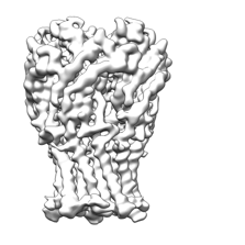
- EMDB-28830: ELIC with Propylamine in saposin nanodiscs with 2:1:1 POPC:POPE:POPG -
+ Open data

- Basic information
| Entry |  | |||||||||
|---|---|---|---|---|---|---|---|---|---|---|
| Title | ELIC with Propylamine in saposin nanodiscs with 2:1:1 POPC:POPE:POPG | |||||||||
 Map data Map data | Propylamine bound Wild Type ELIC in 2:1:1 POPC: POPE: POPG in saposin | |||||||||
 Sample Sample |
| |||||||||
 Keywords Keywords | ELIC /  ion channel / pLGIC / Structural Protein / Membrane Protein / TRANSPORT PROTEIN ion channel / pLGIC / Structural Protein / Membrane Protein / TRANSPORT PROTEIN | |||||||||
| Function / homology |  Function and homology information Function and homology informationextracellular ligand-gated monoatomic ion channel activity / transmembrane signaling receptor activity / membrane / identical protein bindingSimilarity search - Function | |||||||||
| Biological species |  Dickeya dadantii (bacteria) Dickeya dadantii (bacteria) | |||||||||
| Method | single particle reconstruction / cryo EM / Resolution: 3.28 Å | |||||||||
 Authors Authors | Dalal V / Arcario MJ / Petroff II JT / Deitzen NM / Tan BK / Brannigan G / Cheng WWL | |||||||||
| Funding support |  United States, 1 items United States, 1 items
| |||||||||
 Citation Citation | Journal: Nat Commun / Year: 2024 Title: Lipid nanodisc scaffold and size alter the structure of a pentameric ligand-gated ion channel. Authors: Vikram Dalal / Mark J Arcario / John T Petroff / Brandon K Tan / Noah M Dietzen / Michael J Rau / James A J Fitzpatrick / Grace Brannigan / Wayland W L Cheng / Abstract: Lipid nanodiscs have become a standard tool for studying membrane proteins, including using single particle cryo-electron microscopy (cryo-EM). We find that reconstituting the pentameric ligand-gated ...Lipid nanodiscs have become a standard tool for studying membrane proteins, including using single particle cryo-electron microscopy (cryo-EM). We find that reconstituting the pentameric ligand-gated ion channel (pLGIC), Erwinia ligand-gated ion channel (ELIC), in different nanodiscs produces distinct structures by cryo-EM. The effect of the nanodisc on ELIC structure extends to the extracellular domain and agonist binding site. Additionally, molecular dynamic simulations indicate that nanodiscs of different size impact ELIC structure and that the nanodisc scaffold directly interacts with ELIC. These findings suggest that the nanodisc plays a crucial role in determining the structure of pLGICs, and that reconstitution of ion channels in larger nanodiscs may better approximate a lipid membrane environment. | |||||||||
| History |
|
- Structure visualization
| Supplemental images |
|---|
- Downloads & links
-EMDB archive
| Map data | emd_28830.map.gz | 78.1 MB | EMDB map data format | |
|---|---|---|---|---|
| Header (meta data) | emd-28830-v30.xmlemd-28830.xml | 20.1 KB 20.1 KB | Display Display | EMDB header |
| FSC (resolution estimation) | emd_28830_fsc.xml | 10 KB | Display | FSC data file |
| Images |  emd_28830.png emd_28830.png | 34.9 KB | ||
| Filedesc metadata | emd-28830.cif.gz | 6 KB | ||
| Others | emd_28830_additional_1.map.gzemd_28830_half_map_1.map.gzemd_28830_half_map_2.map.gz | 64.4 MB 65.1 MB 65.1 MB | ||
| Archive directory |  http://ftp.pdbj.org/pub/emdb/structures/EMD-28830ftp://ftp.pdbj.org/pub/emdb/structures/EMD-28830 http://ftp.pdbj.org/pub/emdb/structures/EMD-28830ftp://ftp.pdbj.org/pub/emdb/structures/EMD-28830 | HTTPS FTP |
-Related structure data
| Related structure data |  8f33MC  8f32C  8f34C  8f35C  8twvC  8twzC C: citing same article ( M: atomic model generated by this map |
|---|---|
| Similar structure data |


-Links
| EMDB pages | EMDB (EBI/PDBe) / EMDataResource |
|---|---|
| Related items in Molecule of the Month |


-Map
| File | Download / File: emd_28830.map.gz / Format: CCP4 / Size: 83.7 MB / Type: IMAGE STORED AS FLOATING POINT NUMBER (4 BYTES) | ||||||||||||||||||||
|---|---|---|---|---|---|---|---|---|---|---|---|---|---|---|---|---|---|---|---|---|---|
| Annotation | Propylamine bound Wild Type ELIC in 2:1:1 POPC: POPE: POPG in saposin | ||||||||||||||||||||
| Voxel size | X=Y=Z: 0.657 Å | ||||||||||||||||||||
| Density |
| ||||||||||||||||||||
| Symmetry | Space group: 1 | ||||||||||||||||||||
| Details | EMDB XML:
|
-Supplemental data
-Additional map: Propylamine bound Wild Type ELIC in 2:1:1 POPC: POPE: POPG in saposin
| File | emd_28830_additional_1.map | ||||||||||||
|---|---|---|---|---|---|---|---|---|---|---|---|---|---|
| Annotation | Propylamine bound Wild Type ELIC in 2:1:1 POPC: POPE: POPG in saposin | ||||||||||||
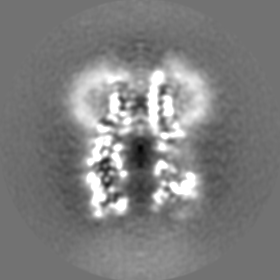
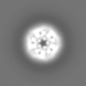
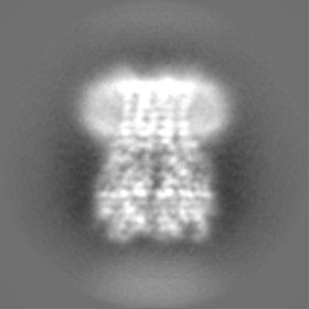
| Projections & Slices |
| ||||||||||||
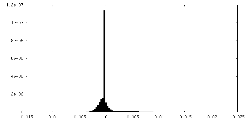
| Density Histograms |
 Z
Z Y
Y X
X


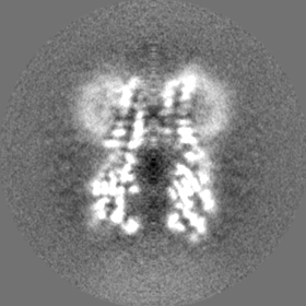
-Half map: Propylamine bound Wild Type ELIC in 2:1:1 POPC: POPE: POPG in saposin
| File | emd_28830_half_map_1.map | ||||||||||||
|---|---|---|---|---|---|---|---|---|---|---|---|---|---|
| Annotation | Propylamine bound Wild Type ELIC in 2:1:1 POPC: POPE: POPG in saposin | ||||||||||||
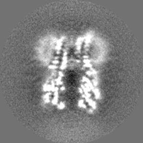
| Projections & Slices |
| ||||||||||||
| Density Histograms |


-Half map: Propylamine bound Wild Type ELIC in 2:1:1 POPC: POPE: POPG in saposin
| File | emd_28830_half_map_2.map | ||||||||||||
|---|---|---|---|---|---|---|---|---|---|---|---|---|---|
| Annotation | Propylamine bound Wild Type ELIC in 2:1:1 POPC: POPE: POPG in saposin | ||||||||||||
| Projections & Slices |
| ||||||||||||
| Density Histograms |


- Sample components
-Entire : ELIC with Propylamine in saposin nanodiscs with 2:1:1 POPC:POPE:POPG
| Entire | Name: ELIC with Propylamine in saposin nanodiscs with 2:1:1 POPC:POPE:POPG |
|---|---|
| Components |
|
-Supramolecule #1: ELIC with Propylamine in saposin nanodiscs with 2:1:1 POPC:POPE:POPG
| Supramolecule | Name: ELIC with Propylamine in saposin nanodiscs with 2:1:1 POPC:POPE:POPG type: complex / ID: 1 / Parent: 0 / Macromolecule list: #1 |
|---|---|
| Source (natural) | Organism: Dickeya dadantii (bacteria) |
-Macromolecule #1: Erwinia chrysanthemi ligand-gated ion channel
| Macromolecule | Name: Erwinia chrysanthemi ligand-gated ion channel / type: protein_or_peptide / ID: 1 / Number of copies: 5 / Enantiomer: LEVO |
|---|---|
| Source (natural) | Organism: Dickeya dadantii (bacteria) |
| Molecular weight | Theoretical: 36.879 KDa |
| Recombinant expression | Organism: Escherichia coli (E. coli) |
| Sequence | String: APADNAADAR PVDVSVSIFI NKIYGVNTLE QTYKVDGYIV AQWTGKPRKT PGDKPLIVEN TQIERWINNG LWVPALEFIN VVGSPDTGN KRLMLFPDGR VIYNARFLGS FSNDMDFRLF PFDRQQFVLE LEPFSYNNQQ LRFSDIQVYT ENIDNEEIDE W WIRGKAST ...String: APADNAADAR PVDVSVSIFI NKIYGVNTLE QTYKVDGYIV AQWTGKPRKT PGDKPLIVEN TQIERWINNG LWVPALEFIN VVGSPDTGN KRLMLFPDGR VIYNARFLGS FSNDMDFRLF PFDRQQFVLE LEPFSYNNQQ LRFSDIQVYT ENIDNEEIDE W WIRGKAST HISDIRYDHL SSVQPNQNEF SRITVRIDAV RNPSYYLWSF ILPLGLIIAA SWSVFWLESF SERLQTSFTL ML TVVAYAF YTSNILPRLP YTTVIDQMII AGYGSIFAAI LLIIFAHHRQ ANGVEDDLLI QRCRLAFPLG FLAIGCVLVI RGI TL UniProtKB: Gamma-aminobutyric-acid receptor subunit beta-1 |
-Macromolecule #2: 3-AMINOPROPANE
| Macromolecule | Name: 3-AMINOPROPANE / type: ligand / ID: 2 / Number of copies: 5 / Formula: 3CN |
|---|---|
| Molecular weight | Theoretical: 59.11 Da |
| Chemical component information | 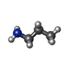 ChemComp-3CN: |
-Experimental details
-Structure determination
| Method | cryo EM |
|---|---|
 Processing Processing | single particle reconstruction |
| Aggregation state | particle |
-Sample preparation
| Buffer | pH: 7.5 |
|---|---|
| Grid | Model: Quantifoil R2/2 / Material: COPPER / Mesh: 300 / Pretreatment - Type: PLASMA CLEANING / Pretreatment - Time: 60 sec. / Pretreatment - Atmosphere: OTHER / Pretreatment - Pressure: 9.332 kPa / Details: O2 27.5 sccm H2 6.4 sccm |
| Vitrification | Cryogen name: ETHANE / Chamber humidity: 100 % / Chamber temperature: 277 K / Instrument: FEI VITROBOT MARK IV / Details: Blot for 2 seconds before plunging. |
- Electron microscopy
| Microscope | FEI TITAN KRIOS |
|---|---|
| Electron beam | Acceleration voltage: 300 kV / Electron source: FIELD EMISSION GUN |
| Electron optics | C2 aperture diameter: 50.0 µm / Calibrated magnification: 96000 / Illumination mode: FLOOD BEAM / Imaging mode: BRIGHT FIELDBright-field microscopy / Cs: 0.01 mm / Nominal defocus max: 2.4 µm / Nominal defocus min: 0.8 µm / Nominal magnification: 96000 |
| Specialist optics | Spherical aberration corrector: Microscope was equipped with a Cs corrector |
| Sample stage | Specimen holder model: FEI TITAN KRIOS AUTOGRID HOLDER / Cooling holder cryogen: NITROGEN |
| Image recording | Film or detector model: FEI FALCON IV (4k x 4k) / Digitization - Dimensions - Width: 4096 pixel / Digitization - Dimensions - Height: 4096 pixel / Number grids imaged: 1 / Average exposure time: 4.28 sec. / Average electron dose: 47.55 e/Å2 |
| Experimental equipment |  Model: Titan Krios / Image courtesy: FEI Company |
-Image processing
| Startup model | Type of model: PDB ENTRY PDB model - PDB ID: |
|---|---|
| Initial angle assignment | Type: ANGULAR RECONSTITUTION |
| Final angle assignment | Type: ANGULAR RECONSTITUTION |
| Final reconstruction | Resolution.type: BY AUTHOR / Resolution: 3.28 Å / Resolution method: FSC 0.143 CUT-OFF / Number images used: 84696 |
| FSC plot (resolution estimation) |  |

-Atomic model buiding 1
| Refinement | Space: REAL / Protocol: AB INITIO MODEL |
|---|---|
| Output model | PDB-8f33: |
