 Movie
Movie Controller


+ Open data

- Basic information
| Entry |  | |||||||||
|---|---|---|---|---|---|---|---|---|---|---|
| Title | Brain-derived 42-residue amyloid-beta fibril type B | |||||||||
 Map data Map data | ||||||||||
 Sample Sample |
| |||||||||
| Function / homology |  Function and homology information Function and homology informationregulation of epidermal growth factor-activated receptor activity / signaling receptor activator activity / collateral sprouting in absence of injury / cytosolic mRNA polyadenylation / microglia development / regulation of synapse structure or activity / Formyl peptide receptors bind formyl peptides and many other ligands / axo-dendritic transport / synaptic assembly at neuromuscular junction / smooth endoplasmic reticulum calcium ion homeostasis ...regulation of epidermal growth factor-activated receptor activity / signaling receptor activator activity / collateral sprouting in absence of injury / cytosolic mRNA polyadenylation / microglia development / regulation of synapse structure or activity / Formyl peptide receptors bind formyl peptides and many other ligands / axo-dendritic transport / synaptic assembly at neuromuscular junction / smooth endoplasmic reticulum calcium ion homeostasis / axon midline choice point recognition / astrocyte activation involved in immune response / regulation of spontaneous synaptic transmission /  regulation of Wnt signaling pathway / mating behavior / ciliary rootlet / Lysosome Vesicle Biogenesis / PTB domain binding / Golgi-associated vesicle / positive regulation of amyloid fibril formation / neuron remodeling / Insertion of tail-anchored proteins into the endoplasmic reticulum membrane / : / Deregulated CDK5 triggers multiple neurodegenerative pathways in Alzheimer's disease models / presynaptic active zone / nuclear envelope lumen / modulation of excitatory postsynaptic potential / suckling behavior / COPII-coated ER to Golgi transport vesicle / dendrite development / smooth endoplasmic reticulum / regulation of NMDA receptor activity / TRAF6 mediated NF-kB activation / Advanced glycosylation endproduct receptor signaling / neuromuscular process controlling balance / regulation of presynapse assembly / The NLRP3 inflammasome / intracellular copper ion homeostasis / transition metal ion binding / regulation of multicellular organism growth / negative regulation of long-term synaptic potentiation / negative regulation of neuron differentiation / ECM proteoglycans / spindle midzone / positive regulation of T cell migration / Purinergic signaling in leishmaniasis infection / positive regulation of calcium-mediated signaling / forebrain development / regulation of peptidyl-tyrosine phosphorylation / positive regulation of chemokine production / clathrin-coated pit / Notch signaling pathway / positive regulation of G2/M transition of mitotic cell cycle / ionotropic glutamate receptor signaling pathway / positive regulation of protein metabolic process / neuron projection maintenance / cholesterol metabolic process / extracellular matrix organization / positive regulation of glycolytic process / positive regulation of mitotic cell cycle / response to interleukin-1 / axonogenesis / adult locomotory behavior / trans-Golgi network membrane / dendritic shaft / locomotory behavior / platelet alpha granule lumen / positive regulation of peptidyl-threonine phosphorylation / learning / central nervous system development / positive regulation of interleukin-1 beta production / positive regulation of long-term synaptic potentiation / astrocyte activation / endosome lumen / synapse organization / Post-translational protein phosphorylation / regulation of long-term neuronal synaptic plasticity / positive regulation of JNK cascade / microglial cell activation / TAK1-dependent IKK and NF-kappa-B activation / visual learning / neuromuscular junction / serine-type endopeptidase inhibitor activity / recycling endosome / cognition / positive regulation of inflammatory response / Golgi lumen / neuron cellular homeostasis / endocytosis / positive regulation of interleukin-6 production / positive regulation of non-canonical NF-kappaB signal transduction / cellular response to amyloid-beta / Regulation of Insulin-like Growth Factor (IGF) transport and uptake by Insulin-like Growth Factor Binding Proteins (IGFBPs) / neuron projection development / positive regulation of DNA-binding transcription factor activity / G2/M transition of mitotic cell cycle / cell-cell junction / synaptic vesicle / positive regulation of tumor necrosis factor production / regulation of translation regulation of Wnt signaling pathway / mating behavior / ciliary rootlet / Lysosome Vesicle Biogenesis / PTB domain binding / Golgi-associated vesicle / positive regulation of amyloid fibril formation / neuron remodeling / Insertion of tail-anchored proteins into the endoplasmic reticulum membrane / : / Deregulated CDK5 triggers multiple neurodegenerative pathways in Alzheimer's disease models / presynaptic active zone / nuclear envelope lumen / modulation of excitatory postsynaptic potential / suckling behavior / COPII-coated ER to Golgi transport vesicle / dendrite development / smooth endoplasmic reticulum / regulation of NMDA receptor activity / TRAF6 mediated NF-kB activation / Advanced glycosylation endproduct receptor signaling / neuromuscular process controlling balance / regulation of presynapse assembly / The NLRP3 inflammasome / intracellular copper ion homeostasis / transition metal ion binding / regulation of multicellular organism growth / negative regulation of long-term synaptic potentiation / negative regulation of neuron differentiation / ECM proteoglycans / spindle midzone / positive regulation of T cell migration / Purinergic signaling in leishmaniasis infection / positive regulation of calcium-mediated signaling / forebrain development / regulation of peptidyl-tyrosine phosphorylation / positive regulation of chemokine production / clathrin-coated pit / Notch signaling pathway / positive regulation of G2/M transition of mitotic cell cycle / ionotropic glutamate receptor signaling pathway / positive regulation of protein metabolic process / neuron projection maintenance / cholesterol metabolic process / extracellular matrix organization / positive regulation of glycolytic process / positive regulation of mitotic cell cycle / response to interleukin-1 / axonogenesis / adult locomotory behavior / trans-Golgi network membrane / dendritic shaft / locomotory behavior / platelet alpha granule lumen / positive regulation of peptidyl-threonine phosphorylation / learning / central nervous system development / positive regulation of interleukin-1 beta production / positive regulation of long-term synaptic potentiation / astrocyte activation / endosome lumen / synapse organization / Post-translational protein phosphorylation / regulation of long-term neuronal synaptic plasticity / positive regulation of JNK cascade / microglial cell activation / TAK1-dependent IKK and NF-kappa-B activation / visual learning / neuromuscular junction / serine-type endopeptidase inhibitor activity / recycling endosome / cognition / positive regulation of inflammatory response / Golgi lumen / neuron cellular homeostasis / endocytosis / positive regulation of interleukin-6 production / positive regulation of non-canonical NF-kappaB signal transduction / cellular response to amyloid-beta / Regulation of Insulin-like Growth Factor (IGF) transport and uptake by Insulin-like Growth Factor Binding Proteins (IGFBPs) / neuron projection development / positive regulation of DNA-binding transcription factor activity / G2/M transition of mitotic cell cycle / cell-cell junction / synaptic vesicle / positive regulation of tumor necrosis factor production / regulation of translationSimilarity search - Function | |||||||||
| Biological species |  Homo sapiens (human) Homo sapiens (human) | |||||||||
| Method | helical reconstruction / cryo EM / Resolution: 2.76 Å | |||||||||
 Authors Authors | Tycko R / Lee M / Yau Y-M / Louis JM | |||||||||
| Funding support |  United States, 1 items United States, 1 items
| |||||||||
 Citation Citation | Journal: Proc Natl Acad Sci U S A / Year: 2023 Title: Structures of brain-derived 42-residue amyloid-β fibril polymorphs with unusual molecular conformations and intermolecular interactions. Authors: Myungwoon Lee / Wai-Ming Yau / John M Louis / Robert Tycko / Abstract: Fibrils formed by the 42-residue amyloid-β peptide (Aβ42), a main component of amyloid deposits in Alzheimer's disease (AD), are known to be polymorphic, i.e., to contain multiple possible ...Fibrils formed by the 42-residue amyloid-β peptide (Aβ42), a main component of amyloid deposits in Alzheimer's disease (AD), are known to be polymorphic, i.e., to contain multiple possible molecular structures. Previous studies of Aβ42 fibrils, including fibrils prepared entirely in vitro or extracted from brain tissue and using solid-state NMR (ssNMR) or cryogenic electron microscopy (cryo-EM) methods, have found polymorphs with differences in amino acid sidechain orientations, lengths of structurally ordered segments, and contacts between cross-β subunit pairs within a single filament. Despite these differences, Aβ42 molecules adopt a common S-shaped conformation in all previously described high-resolution Aβ42 fibril structures. Here we report two cryo-EM-based structures of Aβ42 fibrils that are qualitatively different, in samples derived from AD brain tissue by seeded growth. In type A fibrils, residues 12 to 42 adopt a ν-shaped conformation, with both intra-subunit and intersubunit hydrophobic contacts to form a compact core. In type B fibrils, residues 2 to 42 adopt an υ-shaped conformation, with only intersubunit contacts and internal pores. Type A and type B fibrils have opposite helical handedness. Cryo-EM density maps and molecular dynamics simulations indicate intersubunit K16-A42 salt bridges in type B fibrils and partially occupied K28-A42 salt bridges in type A fibrils. The coexistence of two predominant polymorphs, with differences in N-terminal dynamics, is supported by ssNMR data, as is faithful propagation of structures from first-generation to second-generation brain-seeded Aβ42 fibril samples. These results demonstrate that Aβ42 fibrils can exhibit a greater range of structural variations than seen in previous studies. | |||||||||
| History |
|
- Structure visualization
| Supplemental images |
|---|

- Downloads & links
-EMDB archive
| Map data | emd_28741.map.gz | 25.3 MB | EMDB map data format | |
|---|---|---|---|---|
| Header (meta data) | emd-28741-v30.xmlemd-28741.xml | 17.1 KB 17.1 KB | Display Display | EMDB header |
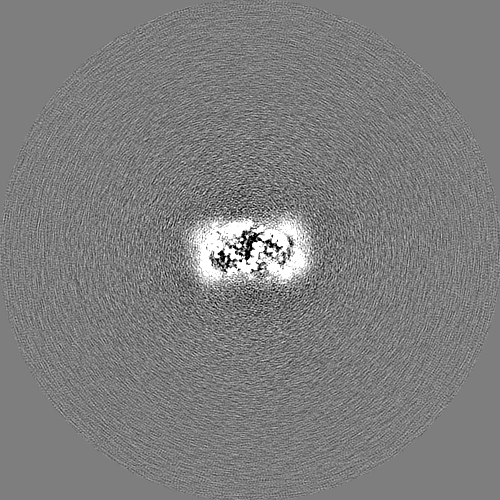
| Images | 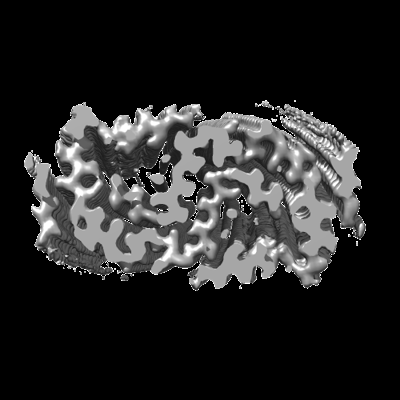 emd_28741.png emd_28741.png | 67.1 KB | ||
| Others | emd_28741_half_map_1.map.gzemd_28741_half_map_2.map.gz | 383.3 MB 383.1 MB | ||
| Archive directory |  http://ftp.pdbj.org/pub/emdb/structures/EMD-28741ftp://ftp.pdbj.org/pub/emdb/structures/EMD-28741 http://ftp.pdbj.org/pub/emdb/structures/EMD-28741ftp://ftp.pdbj.org/pub/emdb/structures/EMD-28741 | HTTPS FTP |
-Related structure data
| Related structure data |  8ezeMC  8ezdC C: citing same article ( M: atomic model generated by this map |
|---|---|
| Similar structure data |
-Links
| EMDB pages | EMDB (EBI/PDBe) / EMDataResource |
|---|---|
| Related items in Molecule of the Month |


-Map
| File | Download / File: emd_28741.map.gz / Format: CCP4 / Size: 476.8 MB / Type: IMAGE STORED AS FLOATING POINT NUMBER (4 BYTES) | ||||||||||||||||||||
|---|---|---|---|---|---|---|---|---|---|---|---|---|---|---|---|---|---|---|---|---|---|
| Voxel size | X=Y=Z: 0.86 Å | ||||||||||||||||||||
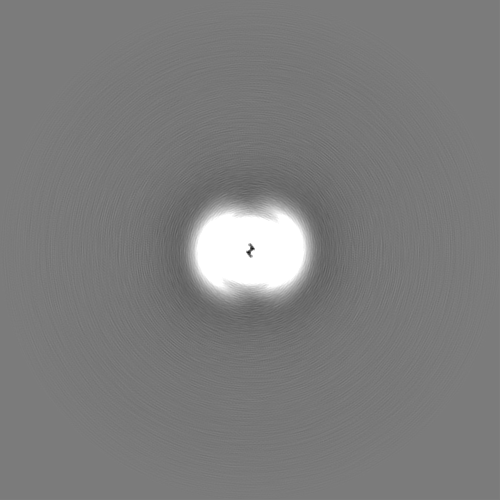
| Density |
| ||||||||||||||||||||
| Symmetry | Space group: 1 | ||||||||||||||||||||
| Details | EMDB XML:
|
-Supplemental data
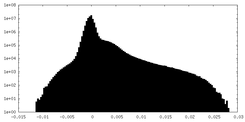
-Half map: #2
| File | emd_28741_half_map_1.map | ||||||||||||
|---|---|---|---|---|---|---|---|---|---|---|---|---|---|
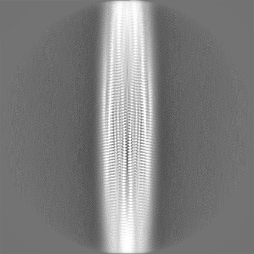
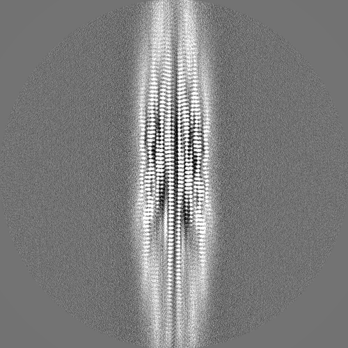
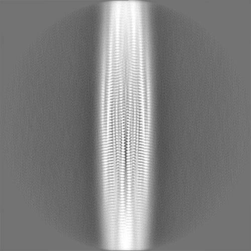
| Projections & Slices |
| ||||||||||||
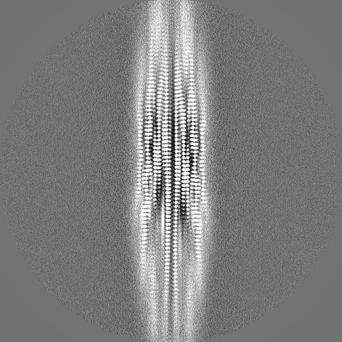
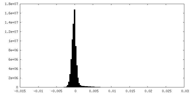
| Density Histograms |
 Z
Z Y
Y X
X


-Half map: #1
| File | emd_28741_half_map_2.map | ||||||||||||
|---|---|---|---|---|---|---|---|---|---|---|---|---|---|
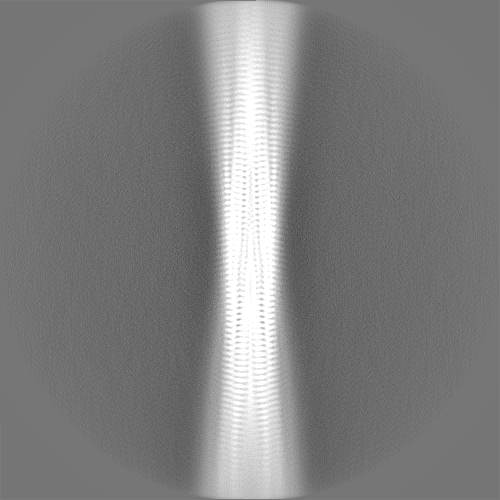
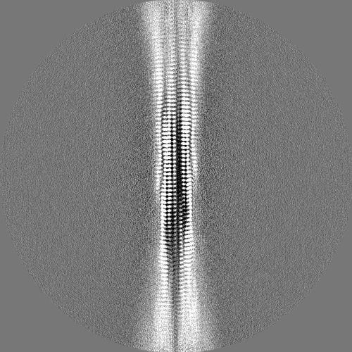
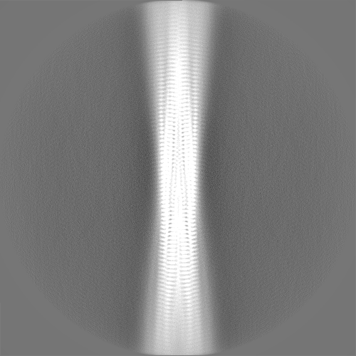
| Projections & Slices |
| ||||||||||||
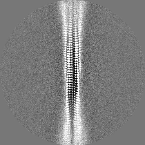
| Density Histograms |

- Sample components
-Entire : amyloid-b 42 (Ab42) fibril
| Entire | Name: amyloid-b 42 (Ab42) fibril |
|---|---|
| Components |
|
-Supramolecule #1: amyloid-b 42 (Ab42) fibril
| Supramolecule | Name: amyloid-b 42 (Ab42) fibril / type: complex / ID: 1 / Chimera: Yes / Parent: 0 / Macromolecule list: all |
|---|---|
| Source (natural) | Organism: Homo sapiens (human) |
| Molecular weight | Theoretical: 4514.10 kDa/nm |
-Macromolecule #1: Beta-amyloid protein 42
| Macromolecule | Name: Beta-amyloid protein 42 / type: protein_or_peptide / ID: 1 / Number of copies: 8 / Enantiomer: LEVO |
|---|---|
| Source (natural) | Organism: Homo sapiens (human) |
| Molecular weight | Theoretical: 4.520087 KDa |
| Recombinant expression | Organism:  Escherichia coli BL21(DE3) (bacteria) Escherichia coli BL21(DE3) (bacteria) |
| Sequence | String: DAEFRHDSGY EVHHQKLVFF AEDVGSNKGA IIGLMVGGVV IA |
-Experimental details
-Structure determination
| Method | cryo EM |
|---|---|
 Processing Processing | helical reconstruction |
| Aggregation state | filament |
-Sample preparation
| Concentration | 0.34 mg/mL |
|---|---|
| Buffer | pH: 7.4 / Component - Concentration: 10.0 mM / Component - Name: Sodium Phosphate / Details: 10mM Na-phosphate, 0.1% sodium azide |
| Grid | Model: Quantifoil R1.2/1.3 / Material: COPPER / Mesh: 400 / Pretreatment - Type: GLOW DISCHARGE / Pretreatment - Time: 45 sec. / Pretreatment - Pressure: 0.039 kPa Details: The grid was glow discharged immediately before use. |
| Vitrification | Cryogen name: ETHANE / Chamber humidity: 99 % / Chamber temperature: 93 K / Instrument: FEI VITROBOT MARK I Details: Preblot for 12-13 seconds and blot for 2.5-3.0 seconds before plunging. |
- Electron microscopy
| Microscope | FEI TITAN KRIOS |
|---|---|
| Electron beam | Acceleration voltage: 300 kV / Electron source: FIELD EMISSION GUN |
| Electron optics | Illumination mode: FLOOD BEAM / Imaging mode: BRIGHT FIELDBright-field microscopy / Nominal defocus max: 2.5 µm / Nominal defocus min: 0.5 µm / Nominal magnification: 130000 |
| Sample stage | Specimen holder model: FEI TITAN KRIOS AUTOGRID HOLDER / Cooling holder cryogen: NITROGEN |
| Image recording | Film or detector model: GATAN K2 SUMMIT (4k x 4k) / Detector mode: SUPER-RESOLUTION / Digitization - Dimensions - Width: 11520 pixel / Digitization - Dimensions - Height: 8184 pixel / Number grids imaged: 1 / Number real images: 5557 / Average exposure time: 1.65 sec. / Average electron dose: 50.34 e/Å2 |
| Experimental equipment |  Model: Titan Krios / Image courtesy: FEI Company |
-Image processing
| Segment selection | Number selected: 285760 / Software - Name: RELION (ver. 4.0.0) |
|---|---|
| Startup model | Type of model: NONE / Details: featureless cylinder |
| Final angle assignment | Type: NOT APPLICABLE / Software - Name: RELION (ver. 4.0.0) Details: 2.48 Angstrom helical rise and 179.36 degree helical twist |
| Final reconstruction | Number classes used: 1 Applied symmetry - Helical parameters - Δz: 2.48 Å Applied symmetry - Helical parameters - Δ&Phi: 179.36 ° Applied symmetry - Helical parameters - Axial symmetry: C1 (asymmetric) Algorithm: FOURIER SPACE / Resolution.type: BY AUTHOR / Resolution: 2.76 Å / Resolution method: FSC 0.143 CUT-OFF / Software - Name: RELION (ver. 4.0.0) Details: 3D refinement and post-processing were performed with 21 (screw) symmetry Number images used: 92019 |
| Details | Gatan Imaging Filter (GIF) Quantum LS |
-Atomic model buiding 1
| Details | Manually generated model was fit into the density using PHENIX and UCSF Chimera. Further refinements were performed using Xplor-NIH. |
|---|---|
| Refinement | Protocol: OTHER |
| Output model | PDB-8eze: |
