National Institutes of Health/National Institute Of Allergy and Infectious Diseases (NIH/NIAID)
United States
Citation
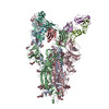
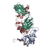
Journal: bioRxiv / Year: 2023 Title: Broadly neutralizing antibody induction by non-stabilized SARS-CoV-2 Spike mRNA vaccination in nonhuman primates. Authors: R Dilshan Malewana / Victoria Stalls / Aaron May / Xiaozhi Lu / David R Martinez / Alexandra Schäfer / Dapeng Li / Maggie Barr / Laura L Sutherland / Esther Lee / Robert Parks / Whitney ...Authors: R Dilshan Malewana / Victoria Stalls / Aaron May / Xiaozhi Lu / David R Martinez / Alexandra Schäfer / Dapeng Li / Maggie Barr / Laura L Sutherland / Esther Lee / Robert Parks / Whitney Edwards Beck / Amanda Newman / Kevin W Bock / Mahnaz Minai / Bianca M Nagata / C Todd DeMarco / Thomas N Denny / Thomas H Oguin / Wes Rountree / Yunfei Wang / Katayoun Mansouri / Robert J Edwards / Gregory D Sempowski / Amanda Eaton / Hiromi Muramatsu / Rory Henderson / Ying Tam / Christopher Barbosa / Juanjie Tang / Derek W Cain / Sampa Santra / Ian N Moore / Hanne Andersen / Mark G Lewis / Hana Golding / Robert Seder / Surender Khurana / David C Montefiori / Norbert Pardi / Drew Weissman / Ralph S Baric / Priyamvada Acharya / Barton F Haynes / Kevin O Saunders / Abstract: Immunization with mRNA or viral vectors encoding spike with diproline substitutions (S-2P) has provided protective immunity against severe COVID-19 disease. How immunization with Severe Acute ...Immunization with mRNA or viral vectors encoding spike with diproline substitutions (S-2P) has provided protective immunity against severe COVID-19 disease. How immunization with Severe Acute Respiratory Syndrome Coronavirus 2 (SARS-CoV-2) spike elicits neutralizing antibodies (nAbs) against difficult-to-neutralize variants of concern (VOCs) remains an area of great interest. Here, we compare immunization of macaques with mRNA vaccines expressing ancestral spike either including or lacking diproline substitutions, and show the diproline substitutions were not required for protection against SARS-CoV-2 challenge or induction of broadly neutralizing B cell lineages. One group of nAbs elicited by the ancestral spike lacking diproline substitutions targeted the outer face of the receptor binding domain (RBD), neutralized all tested SARS-CoV-2 VOCs including Omicron XBB.1.5, but lacked cross-Sarbecovirus neutralization. Structural analysis showed that the macaque broad SARS-CoV-2 VOC nAbs bound to the same epitope as a human broad SARS-CoV-2 VOC nAb, DH1193. Vaccine-induced antibodies that targeted the RBD inner face neutralized multiple Sarbecoviruses, protected mice from bat CoV RsSHC014 challenge, but lacked Omicron variant neutralization. Thus, ancestral SARS-CoV-2 spike lacking proline substitutions encoded by nucleoside-modified mRNA can induce B cell lineages binding to distinct RBD sites that either broadly neutralize animal and human Sarbecoviruses or recent Omicron VOCs.
In the structure databanks used in Yorodumi, some data are registered as the other names, "COVID-19 virus" and "2019-nCoV". Here are the details of the virus and the list of structure data.
Jan 31, 2019. EMDB accession codes are about to change! (news from PDBe EMDB page)
EMDB accession codes are about to change! (news from PDBe EMDB page)
The allocation of 4 digits for EMDB accession codes will soon come to an end. Whilst these codes will remain in use, new EMDB accession codes will include an additional digit and will expand incrementally as the available range of codes is exhausted. The current 4-digit format prefixed with “EMD-” (i.e. EMD-XXXX) will advance to a 5-digit format (i.e. EMD-XXXXX), and so on. It is currently estimated that the 4-digit codes will be depleted around Spring 2019, at which point the 5-digit format will come into force.
The EM Navigator/Yorodumi systems omit the EMD- prefix.
Related info.:Q: What is EMD? / ID/Accession-code notation in Yorodumi/EM Navigator
Yorodumi is a browser for structure data from EMDB, PDB, SASBDB, etc.
This page is also the successor to EM Navigator detail page, and also detail information page/front-end page for Omokage search.
The word "yorodu" (or yorozu) is an old Japanese word meaning "ten thousand". "mi" (miru) is to see.
Related info.:EMDB / PDB / SASBDB / Comparison of 3 databanks / Yorodumi Search / Aug 31, 2016. New EM Navigator & Yorodumi / Yorodumi Papers / Jmol/JSmol / Function and homology information / Changes in new EM Navigator and Yorodumi
 Movie
Movie Controller

 Yorodumi
 Open data
 Basic information
 Map data
 Sample
 Keywords
 Fab /
Fab /  Function and homology information


 Authors
 United States, 1 items
  Citation
 Structure visualization
 Downloads & links
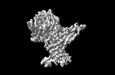 emd_27703.png
 http://ftp.pdbj.org/pub/emdb/structures/EMD-27703
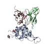


 Z
Z Y
Y X
X


 Sample components
 Processing
 Electron microscopy