Journal: Cell / Year: 2023 Title: Chromatin remodeling of histone H3 variants by DDM1 underlies epigenetic inheritance of DNA methylation. Authors: Seung Cho Lee / Dexter W Adams / Jonathan J Ipsaro / Jonathan Cahn / Jason Lynn / Hyun-Soo Kim / Benjamin Berube / Viktoria Major / Joseph P Calarco / Chantal LeBlanc / Sonali Bhattacharjee ...Authors: Seung Cho Lee / Dexter W Adams / Jonathan J Ipsaro / Jonathan Cahn / Jason Lynn / Hyun-Soo Kim / Benjamin Berube / Viktoria Major / Joseph P Calarco / Chantal LeBlanc / Sonali Bhattacharjee / Umamaheswari Ramu / Daniel Grimanelli / Yannick Jacob / Philipp Voigt / Leemor Joshua-Tor / Robert A Martienssen / Abstract: Nucleosomes block access to DNA methyltransferase, unless they are remodeled by DECREASE in DNA METHYLATION 1 (DDM1), a Snf2-like master regulator of epigenetic inheritance. We show that DDM1 ...Nucleosomes block access to DNA methyltransferase, unless they are remodeled by DECREASE in DNA METHYLATION 1 (DDM1), a Snf2-like master regulator of epigenetic inheritance. We show that DDM1 promotes replacement of histone variant H3.3 by H3.1. In ddm1 mutants, DNA methylation is partly restored by loss of the H3.3 chaperone HIRA, while the H3.1 chaperone CAF-1 becomes essential. The single-particle cryo-EM structure at 3.2 Å of DDM1 with a variant nucleosome reveals engagement with histone H3.3 near residues required for assembly and with the unmodified H4 tail. An N-terminal autoinhibitory domain inhibits activity, while a disulfide bond in the helicase domain supports activity. DDM1 co-localizes with H3.1 and H3.3 during the cell cycle, and with the DNA methyltransferase MET1, but is blocked by H4K16 acetylation. The male germline H3.3 variant MGH3/HTR10 is resistant to remodeling by DDM1 and acts as a placeholder nucleosome in sperm cells for epigenetic inheritance.
Entire : Arabidopsis DDM1 bound to nucleosome (H2A.W, H2B, H3.3, H4, with ...
Entire
Name: Arabidopsis DDM1 bound to nucleosome (H2A.W, H2B, H3.3, H4, with 147 bp DNA)
Components
Complex: Arabidopsis DDM1 bound to nucleosome (H2A.W, H2B, H3.3, H4, with 147 bp DNA)
Complex: DDM1
Protein or peptide: ATP-dependent DNA helicase DDM1
Complex: Nucleosome core particleNucleosome
Complex: Histone octamer
Protein or peptide: Probable histone H2A.7
Protein or peptide: Histone H2B
Protein or peptide: Histone H3.3H3F3A
Protein or peptide: Histone H4
Complex: DNA double helixNucleic acid double helix
DNA: DNA (sense strand)
DNA: DNA (antisense strand)
+
Supramolecule #1: Arabidopsis DDM1 bound to nucleosome (H2A.W, H2B, H3.3, H4, with ...
Supramolecule
Name: Arabidopsis DDM1 bound to nucleosome (H2A.W, H2B, H3.3, H4, with 147 bp DNA) type: complex / ID: 1 / Parent: 0 / Macromolecule list: all Details: Complex contains two copies of each histone (H2A.W, H2B, H3.3, and H4), 147 base pairs of DNA, and 1 copy of Arabidopsis DDM1
Organism: Arabidopsis thaliana (thale cress) / Location in cell: nucleus
+
Supramolecule #3: Nucleosome core particle
Supramolecule
Name: Nucleosome core particle / type: complex / ID: 3 / Parent: 1 / Macromolecule list: #1-#4, #6-#7 Details: Complex contains two copies of each histone (H2A.W, H2B, H3.3, and H4) and 147 base pairs of DNA
+
Supramolecule #4: Histone octamer
Supramolecule
Name: Histone octamer / type: complex / ID: 4 / Parent: 3 / Macromolecule list: #1-#4 Details: Contains two copies each of H2A.W, H2B, H3.3, and H4
Source (natural)
Organism: Arabidopsis thaliana (thale cress) / Location in cell: nucleus
Model: Quantifoil R0.6/1 / Material: COPPER / Support film - Material: CARBON / Support film - topology: HOLEY / Pretreatment - Type: GLOW DISCHARGE / Pretreatment - Time: 70 sec. / Pretreatment - Atmosphere: AIR / Pretreatment - Pressure: 42.0 kPa
Vitrification
Cryogen name: ETHANE / Chamber humidity: 95 % / Chamber temperature: 298 K / Instrument: LEICA EM GP / Details: Blotted for 2.5 seconds before plunging..
Details
DDM1 and reconstituted nucleosomes were reconstituted in a 4:1 molar ratio. Crosslinking with 0.05% glutaraldehyde was performed for 15 minutes followed by quenching with 2 mM Tris, pH 8.0. ATP-gamma-S and MgCl2 were added at 1 and 2 mM, respectively.
-
Electron microscopy
Microscope
TFS KRIOS
Electron beam
Acceleration voltage: 300 kV / Electron source: FIELD EMISSION GUN
Film or detector model: GATAN K3 BIOQUANTUM (6k x 4k) / Detector mode: COUNTING / Number grids imaged: 1 / Number real images: 8165 / Average exposure time: 4.8 sec. / Average electron dose: 71.2 e/Å2
Experimental equipment
Model: Titan Krios / Image courtesy: FEI Company
-
Image processing
Particle selection
Number selected: 3788872
Startup model
Type of model: NONE
Initial angle assignment
Type: MAXIMUM LIKELIHOOD / Software - Name: cryoSPARC (ver. 3.0.2+210831)
Final 3D classification
Number classes: 4 / Avg.num./class: 141299 / Software - Name: cryoSPARC (ver. 3.0.2+210831) Details: Iterative rounds of 2D classification, 3D classification, and subselection were made. The last round of 3D classification had 4 classes of which 1 was chosen for further refinement and final reconstruction.
Final angle assignment
Type: MAXIMUM LIKELIHOOD / Software - Name: cryoSPARC (ver. 3.0.2+210831)
Final reconstruction
Number classes used: 1 / Resolution.type: BY AUTHOR / Resolution: 3.2 Å / Resolution method: FSC 0.143 CUT-OFF / Software - Name: cryoSPARC (ver. 3.0.2+210831) / Number images used: 215066
In the structure databanks used in Yorodumi, some data are registered as the other names, "COVID-19 virus" and "2019-nCoV". Here are the details of the virus and the list of structure data.
Jan 31, 2019. EMDB accession codes are about to change! (news from PDBe EMDB page)
EMDB accession codes are about to change! (news from PDBe EMDB page)
The allocation of 4 digits for EMDB accession codes will soon come to an end. Whilst these codes will remain in use, new EMDB accession codes will include an additional digit and will expand incrementally as the available range of codes is exhausted. The current 4-digit format prefixed with “EMD-” (i.e. EMD-XXXX) will advance to a 5-digit format (i.e. EMD-XXXXX), and so on. It is currently estimated that the 4-digit codes will be depleted around Spring 2019, at which point the 5-digit format will come into force.
The EM Navigator/Yorodumi systems omit the EMD- prefix.
Related info.:Q: What is EMD? / ID/Accession-code notation in Yorodumi/EM Navigator
Yorodumi is a browser for structure data from EMDB, PDB, SASBDB, etc.
This page is also the successor to EM Navigator detail page, and also detail information page/front-end page for Omokage search.
The word "yorodu" (or yorozu) is an old Japanese word meaning "ten thousand". "mi" (miru) is to see.
Related info.:EMDB / PDB / SASBDB / Comparison of 3 databanks / Yorodumi Search / Aug 31, 2016. New EM Navigator & Yorodumi / Yorodumi Papers / Jmol/JSmol / Function and homology information / Changes in new EM Navigator and Yorodumi
 Movie
Movie Controller

 Yorodumi
 Open data
 Basic information
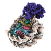
 Map data
 Sample
 Keywords
 Helicase /
  Function and homology information
 Authors
 United States, 1 items
  Citation

 Structure visualization
 Downloads & links
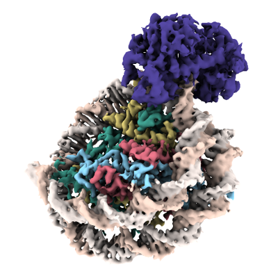 emd_26855.png
 http://ftp.pdbj.org/pub/emdb/structures/EMD-26855
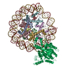


 Z
Z Y
Y X
X


 Sample components
 Processing
 Electron microscopy


