 Movie
Movie Controller

[English] 日本語
 Yorodumi
- EMDB-26728: PARL-cleaved Skd3 (human ClpB) E455Q dodecamer bound to ATPgammaS -
+ Open data

- Basic information
| Entry | 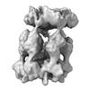 | |||||||||||||||||||||
|---|---|---|---|---|---|---|---|---|---|---|---|---|---|---|---|---|---|---|---|---|---|---|
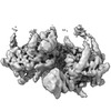
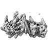
| Title | PARL-cleaved Skd3 (human ClpB) E455Q dodecamer bound to ATPgammaS | |||||||||||||||||||||
 Map data Map data | PARL-cleaved Skd3 (human ClpB) E455Q Nucleotide Binding Domain in the dodecameric state, half map 2 | |||||||||||||||||||||
 Sample Sample |
| |||||||||||||||||||||
 Keywords Keywords | AAA+ ATPase / Chaperone /  Mitochondria / Protein Folding Mitochondria / Protein Folding | |||||||||||||||||||||
| Biological species |  Homo sapiens (human) Homo sapiens (human) | |||||||||||||||||||||
| Method | single particle reconstruction / cryo EM / Resolution: 6.8 Å | |||||||||||||||||||||
 Authors Authors | Gupta A / Lentzsch AM / Siegel AS / Yu Z / Lu C / Chio US / Cheng Y / Shan S | |||||||||||||||||||||
| Funding support |  United States, 6 items United States, 6 items
| |||||||||||||||||||||
 Citation Citation | Journal: Sci Adv / Year: 2023 Title: Dodecamer assembly of a metazoan AAA chaperone couples substrate extraction to refolding. Authors: Arpit Gupta / Alfred M Lentzsch / Alex Siegel / Zanlin Yu / Un Seng Chio / Yifan Cheng / Shu-Ou Shan / Abstract: Ring-forming AAA chaperones solubilize protein aggregates and protect organisms from proteostatic stress. In metazoans, the AAA chaperone Skd3 in the mitochondrial intermembrane space (IMS) is ...Ring-forming AAA chaperones solubilize protein aggregates and protect organisms from proteostatic stress. In metazoans, the AAA chaperone Skd3 in the mitochondrial intermembrane space (IMS) is critical for human health and efficiently refolds aggregated proteins, but its underlying mechanism is poorly understood. Here, we show that Skd3 harbors both disaggregase and protein refolding activities enabled by distinct assembly states. High-resolution structures of Skd3 hexamers in distinct conformations capture ratchet-like motions that mediate substrate extraction. Unlike previously described disaggregases, Skd3 hexamers further assemble into dodecameric cages in which solubilized substrate proteins can attain near-native states. Skd3 mutants defective in dodecamer assembly retain disaggregase activity but are impaired in client refolding, linking the disaggregase and refolding activities to the hexameric and dodecameric states of Skd3, respectively. We suggest that Skd3 is a combined disaggregase and foldase, and this property is particularly suited to meet the complex proteostatic demands in the mitochondrial IMS. | |||||||||||||||||||||
| History |
|
- Structure visualization
| Supplemental images |
|---|

- Downloads & links
-EMDB archive
| Map data | emd_26728.map.gz | 70.8 MB |  EMDB map data format EMDB map data format | |
|---|---|---|---|---|
| Header (meta data) | emd-26728-v30.xmlemd-26728.xml | 16 KB 16 KB | Display Display | EMDB header |
| FSC (resolution estimation) | emd_26728_fsc.xml | 376.7 KB | Display | FSC data file |
| Images |  emd_26728.png emd_26728.png | 75.8 KB | ||
| Filedesc metadata | emd-26728.cif.gz | 5 KB | ||
| Others | emd_26728_half_map_1.map.gzemd_26728_half_map_2.map.gz | 71.1 MB 71.3 MB | ||
| Archive directory |  http://ftp.pdbj.org/pub/emdb/structures/EMD-26728ftp://ftp.pdbj.org/pub/emdb/structures/EMD-26728 http://ftp.pdbj.org/pub/emdb/structures/EMD-26728ftp://ftp.pdbj.org/pub/emdb/structures/EMD-26728 | HTTPS FTP |
-Related structure data


-Links
| EMDB pages | EMDB (EBI/PDBe) / EMDataResource |
|---|
-Map
| File | Download / File: emd_26728.map.gz / Format: CCP4 / Size: 91.1 MB / Type: IMAGE STORED AS FLOATING POINT NUMBER (4 BYTES) | ||||||||||||||||||||||||||||||||||||
|---|---|---|---|---|---|---|---|---|---|---|---|---|---|---|---|---|---|---|---|---|---|---|---|---|---|---|---|---|---|---|---|---|---|---|---|---|---|
| Annotation | PARL-cleaved Skd3 (human ClpB) E455Q Nucleotide Binding Domain in the dodecameric state, half map 2 | ||||||||||||||||||||||||||||||||||||
| Projections & slices | Image control
Images are generated by Spider. | ||||||||||||||||||||||||||||||||||||
| Voxel size | X=Y=Z: 1.3904 Å | ||||||||||||||||||||||||||||||||||||
| Density |
| ||||||||||||||||||||||||||||||||||||
| Symmetry | Space group: 1 | ||||||||||||||||||||||||||||||||||||
| Details | EMDB XML:
|
 Z (Sec.)
 Y (Row.)
 X (Col.)


-Supplemental data
-Half map: PARL-cleaved Skd3 (human ClpB) E455Q Nucleotide Binding Domain...
| File | emd_26728_half_map_1.map | ||||||||||||
|---|---|---|---|---|---|---|---|---|---|---|---|---|---|
| Annotation | PARL-cleaved Skd3 (human ClpB) E455Q Nucleotide Binding Domain in the dodecameric state, half map 1 | ||||||||||||
| Projections & Slices |
| ||||||||||||
| Density Histograms |


-Half map: PARL-cleaved Skd3 (human ClpB) E455Q Nucleotide Binding Domain...
| File | emd_26728_half_map_2.map | ||||||||||||
|---|---|---|---|---|---|---|---|---|---|---|---|---|---|
| Annotation | PARL-cleaved Skd3 (human ClpB) E455Q Nucleotide Binding Domain in the dodecameric state, half map 1 | ||||||||||||
| Projections & Slices |
| ||||||||||||
| Density Histograms |


- Sample components
-Entire : PARL-cleaved Skd3
| Entire | Name: PARL-cleaved Skd3 |
|---|---|
| Components |
|
-Supramolecule #1: PARL-cleaved Skd3
| Supramolecule | Name: PARL-cleaved Skd3 / type: complex / ID: 1 / Parent: 0 / Macromolecule list: all |
|---|---|
| Source (natural) | Organism: Homo sapiens (human) |
| Molecular weight | Theoretical: 792 KDa |
-Macromolecule #1: PARL-cleaved Skd3
| Macromolecule | Name: PARL-cleaved Skd3 / type: protein_or_peptide / ID: 1 / Enantiomer: LEVO |
|---|---|
| Source (natural) | Organism: Homo sapiens (human) |
| Recombinant expression | Organism:  Escherichia coli (E. coli) Escherichia coli (E. coli) |
| Sequence | String: YSKSPSNKDA ALLEAARANN MQEVSRLLSE GADVNAKHRL GWTALMVAAI NRNNSVVQVL LAAGADPNLG DDFSSVYKTA KEQGIHSLED GGQDGASRHI TNQWTSALEF RRWLGLPAGV LITREDDFNN RLNNRASFKG CTALHYAVLA DDYRTVKELL DGGANPLQRN ...String: YSKSPSNKDA ALLEAARANN MQEVSRLLSE GADVNAKHRL GWTALMVAAI NRNNSVVQVL LAAGADPNLG DDFSSVYKTA KEQGIHSLED GGQDGASRHI TNQWTSALEF RRWLGLPAGV LITREDDFNN RLNNRASFKG CTALHYAVLA DDYRTVKELL DGGANPLQRN EMGHTPLDYA REGEVMKLLR TSEAKYQEKQ RKREAEERRR FPLEQRLKEH IIGQESAIAT VGAAIRRKEN GWYDEEHPLV FLFLGSSGIG KTELAKQTAK YMHKDAKKGF IRLDMSEFQE RHEVAKFIGS PPGYVGHEEG GQLTKKLKQC PNAVVLFDQV DKAHPDVLTI MLQLFDEGRL TDGKGKTIDC KDAIFIMTSN VASDEIAQHA LQLRQEALEM SRNRIAENLG DVQISDKITI SKNFKENVIR PILKAHFRRD EFLGRINEIV YFLPFCHSEL IQLVNKELNF WAKRAKQRHN ITLLWDREVA DVLVDGYNVH YGARSIKHEV ERRVVNQLAA AYEQDLLPGG CTLRITVEDS DKQLLKSPEL PSPQAEKRLP KLRLEIIDKD SKTRRLDIRA PLHPEKVCNT I |
-Experimental details
-Structure determination
| Method | cryo EM |
|---|---|
 Processing Processing | single particle reconstruction |
| Aggregation state | particle |
-Sample preparation
| Concentration | 0.1 mg/mL |
|---|---|
| Buffer | pH: 7.4 |
| Vitrification | Cryogen name: ETHANE / Chamber humidity: 100 % |
- Electron microscopy
| Microscope | FEI TITAN KRIOS |
|---|---|
| Electron beam | Acceleration voltage: 300 kV / Electron source: FIELD EMISSION GUN |
| Electron optics | Illumination mode: FLOOD BEAM / Imaging mode: BRIGHT FIELDBright-field microscopy / Nominal defocus max: -2.0 µm / Nominal defocus min: -1.0 µm |
| Image recording | Film or detector model: FEI FALCON IV (4k x 4k) / Average electron dose: 60.0 e/Å2 |
| Experimental equipment |  Model: Titan Krios / Image courtesy: FEI Company |
-Image processing
| Startup model | Type of model: NONE |
|---|---|
| Initial angle assignment | Type: ANGULAR RECONSTITUTION |
| Final angle assignment | Type: ANGULAR RECONSTITUTION |
| Final reconstruction | Resolution.type: BY AUTHOR / Resolution: 6.8 Å / Resolution method: FSC 0.143 CUT-OFF / Number images used: 521193 |
| FSC plot (resolution estimation) |  |
