 Movie
Movie Controller


+ Open data

- Basic information
| Entry |  | |||||||||
|---|---|---|---|---|---|---|---|---|---|---|
| Title | In situ map of the clathrin hub | |||||||||
 Map data Map data | In situ map of the clathrin hub | |||||||||
 Sample Sample |
| |||||||||
 Keywords Keywords |  Clathrin / Actin / ENDOCYTOSIS Clathrin / Actin / ENDOCYTOSIS | |||||||||
| Biological species |  Homo sapiens (human) Homo sapiens (human) | |||||||||
| Method | subtomogram averaging / cryo EM / Resolution: 27.0 Å | |||||||||
 Authors Authors | Serwas D / Akamatsu M / Moayed A / Vegesna K / Vasan R / Hill JM / Schoeneberg J / Davies KM / Rangamani P / Drubin DG | |||||||||
| Funding support |  United States, United States,  France, 2 items France, 2 items
| |||||||||
 Citation Citation | Journal: Dev Cell / Year: 2022 Title: Mechanistic insights into actin force generation during vesicle formation from cryo-electron tomography. Authors: Daniel Serwas / Matthew Akamatsu / Amir Moayed / Karthik Vegesna / Ritvik Vasan / Jennifer M Hill / Johannes Schöneberg / Karen M Davies / Padmini Rangamani / David G Drubin / Abstract: Actin assembly provides force for a multitude of cellular processes. Compared to actin-assembly-based force production during cell migration, relatively little is understood about how actin assembly ...Actin assembly provides force for a multitude of cellular processes. Compared to actin-assembly-based force production during cell migration, relatively little is understood about how actin assembly generates pulling forces for vesicle formation. Here, cryo-electron tomography identified actin filament number, organization, and orientation during clathrin-mediated endocytosis in human SK-MEL-2 cells, showing that force generation is robust despite variance in network organization. Actin dynamics simulations incorporating a measured branch angle indicate that sufficient force to drive membrane internalization is generated through polymerization and that assembly is triggered from ∼4 founding "mother" filaments, consistent with tomography data. Hip1R actin filament anchoring points are present along the entire endocytic invagination, where simulations show that it is key to pulling force generation, and along the neck, where it targets filament growth and makes internalization more robust. Actin organization described here allowed direct translation of structure to mechanism with broad implications for other actin-driven processes. | |||||||||
| History |
|
- Structure visualization
| Supplemental images |
|---|

- Downloads & links
-EMDB archive
| Map data | emd_26484.map.gz | 1.8 MB |  EMDB map data format EMDB map data format | |
|---|---|---|---|---|
| Header (meta data) | emd-26484-v30.xmlemd-26484.xml | 13.5 KB 13.5 KB | Display Display | EMDB header |
| Images | 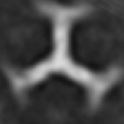 emd_26484.png emd_26484.png | 42.8 KB | ||
| Filedesc metadata | emd-26484.cif.gz | 4.4 KB | ||
| Others | emd_26484_half_map_1.map.gzemd_26484_half_map_2.map.gz | 469.9 KB 467.3 KB | ||
| Archive directory |  http://ftp.pdbj.org/pub/emdb/structures/EMD-26484ftp://ftp.pdbj.org/pub/emdb/structures/EMD-26484 http://ftp.pdbj.org/pub/emdb/structures/EMD-26484ftp://ftp.pdbj.org/pub/emdb/structures/EMD-26484 | HTTPS FTP |
-Related structure data
-Links
| EMDB pages | EMDB (EBI/PDBe) / EMDataResource |
|---|
-Map
| File | Download / File: emd_26484.map.gz / Format: CCP4 / Size: 2.3 MB / Type: IMAGE STORED AS FLOATING POINT NUMBER (4 BYTES) | ||||||||||||||||||||
|---|---|---|---|---|---|---|---|---|---|---|---|---|---|---|---|---|---|---|---|---|---|
| Annotation | In situ map of the clathrin hub | ||||||||||||||||||||
| Voxel size | X=Y=Z: 5.943 Å | ||||||||||||||||||||
| Density |
| ||||||||||||||||||||
| Symmetry | Space group: 1 | ||||||||||||||||||||
| Details | EMDB XML:
|
-Supplemental data
-Half map: Half Map 1
| File | emd_26484_half_map_1.map | ||||||||||||
|---|---|---|---|---|---|---|---|---|---|---|---|---|---|
| Annotation | Half Map 1 | ||||||||||||
| Projections & Slices |
| ||||||||||||
| Density Histograms |
 Z
Z Y
Y X
X


-Half map: Half Map 2
| File | emd_26484_half_map_2.map | ||||||||||||
|---|---|---|---|---|---|---|---|---|---|---|---|---|---|
| Annotation | Half Map 2 | ||||||||||||
| Projections & Slices |
| ||||||||||||
| Density Histograms |


- Sample components
-Entire : In situ map of the clathrin hub
| Entire | Name: In situ map of the clathrin hub |
|---|---|
| Components |
|
-Supramolecule #1: In situ map of the clathrin hub
| Supramolecule | Name: In situ map of the clathrin hub / type: cell / ID: 1 / Parent: 0 |
|---|---|
| Source (natural) | Organism: Homo sapiens (human) / Strain: SK-MEL-2 |
-Experimental details
-Structure determination
| Method | cryo EM |
|---|---|
 Processing Processing | subtomogram averaging |
| Aggregation state | cell |
-Sample preparation
| Buffer | pH: 7.4 |
|---|---|
| Grid | Model: Quantifoil / Material: GOLD / Mesh: 200 / Support film - Material: CARBON / Support film - topology: HOLEY |
| Vitrification | Cryogen name: ETHANE |
- Electron microscopy
| Microscope | FEI TITAN KRIOS |
|---|---|
| Electron beam | Acceleration voltage: 300 kV / Electron source: FIELD EMISSION GUN |
| Electron optics | Illumination mode: FLOOD BEAM / Imaging mode: BRIGHT FIELDBright-field microscopy / Nominal defocus max: -0.0152562 µm / Nominal defocus min: 0.0052084 µm |
| Image recording | Film or detector model: GATAN K2 SUMMIT (4k x 4k) / Detector mode: SUPER-RESOLUTION / Average electron dose: 2.5 e/Å2 |
| Experimental equipment |  Model: Titan Krios / Image courtesy: FEI Company |
-Image processing
| Extraction | Number tomograms: 7 / Number images used: 561 |
|---|---|
| Final angle assignment | Type: NOT APPLICABLE |
| Final reconstruction | Applied symmetry - Point group: C3 (3 fold cyclic) / Algorithm: BACK PROJECTION / Resolution.type: BY AUTHOR / Resolution: 27.0 Å / Resolution method: FSC 0.5 CUT-OFF / Software: (Name: IMOD, PEET) Details: Half maps were generated using the fnHalfVolume function of calcFSC in PEET rather than "gold standard" FSC. Number subtomograms used: 472 |
