 Movie
Movie Controller


+ Open data

- Basic information
| Entry |  | |||||||||
|---|---|---|---|---|---|---|---|---|---|---|
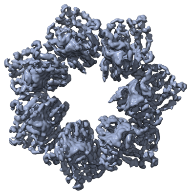
| Title | Heptameric Human Twinkle Helicase Clinical Variant W315L | |||||||||
 Map data Map data | ||||||||||
 Sample Sample |
| |||||||||
 Keywords Keywords |  Helicase / walker A / walker B / DNA binding / DNA BINDING PROTEIN Helicase / walker A / walker B / DNA binding / DNA BINDING PROTEIN | |||||||||
| Function / homology |  Function and homology information Function and homology informationDNA 5'-3' helicase / mitochondrial DNA replication / mitochondrial transcription / 5'-3' DNA helicase activity / protein hexamerization / DNA unwinding involved in DNA replication / mitochondrial nucleoid / isomerase activity / DNA helicase activity / cellular response to glucose stimulus ...DNA 5'-3' helicase / mitochondrial DNA replication / mitochondrial transcription / 5'-3' DNA helicase activity / protein hexamerization / DNA unwinding involved in DNA replication / mitochondrial nucleoid / isomerase activity / DNA helicase activity / cellular response to glucose stimulus / Transcriptional activation of mitochondrial biogenesis / single-stranded DNA binding / mitochondrial inner membrane / protease binding / mitochondrial matrix / lipid binding / ATP hydrolysis activity / mitochondrion / ATP binding / identical protein bindingSimilarity search - Function | |||||||||
| Biological species |  Homo sapiens (human) Homo sapiens (human) | |||||||||
| Method | single particle reconstruction / cryo EM / Resolution: 4.5 Å | |||||||||
 Authors Authors | Riccio AA / Bouvette J | |||||||||
| Funding support |  United States, 2 items United States, 2 items
| |||||||||
 Citation Citation | Journal: Proc Natl Acad Sci U S A / Year: 2022 Title: Structural insight and characterization of human Twinkle helicase in mitochondrial disease. Authors: Amanda A Riccio / Jonathan Bouvette / Lalith Perera / Matthew J Longley / Juno M Krahn / Jason G Williams / Robert Dutcher / Mario J Borgnia / William C Copeland / Abstract: Twinkle is the mammalian helicase vital for replication and integrity of mitochondrial DNA. Over 90 Twinkle helicase disease variants have been linked to progressive external ophthalmoplegia and ...Twinkle is the mammalian helicase vital for replication and integrity of mitochondrial DNA. Over 90 Twinkle helicase disease variants have been linked to progressive external ophthalmoplegia and ataxia neuropathies among other mitochondrial diseases. Despite the biological and clinical importance, Twinkle represents the only remaining component of the human minimal mitochondrial replisome that has yet to be structurally characterized. Here, we present 3-dimensional structures of human Twinkle W315L. Employing cryo-electron microscopy (cryo-EM), we characterize the oligomeric assemblies of human full-length Twinkle W315L, define its multimeric interface, and map clinical variants associated with Twinkle in inherited mitochondrial disease. Cryo-EM, crosslinking-mass spectrometry, and molecular dynamics simulations provide insight into the dynamic movement and molecular consequences of the W315L clinical variant. Collectively, this ensemble of structures outlines a framework for studying Twinkle function in mitochondrial DNA replication and associated disease states. | |||||||||
| History |
|
- Structure visualization
| Supplemental images |
|---|
- Downloads & links
-EMDB archive
| Map data | emd_25744.map.gz | 218.2 MB | EMDB map data format | |
|---|---|---|---|---|
| Header (meta data) | emd-25744-v30.xmlemd-25744.xml | 14.8 KB 14.8 KB | Display Display | EMDB header |
| FSC (resolution estimation) | emd_25744_fsc.xml | 13.3 KB | Display | FSC data file |
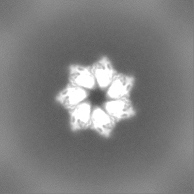
| Images |  emd_25744.png emd_25744.png | 180.8 KB | ||
| Masks | emd_25744_msk_1.map | 244.1 MB | Mask map | |
| Filedesc metadata | emd-25744.cif.gz | 5.6 KB | ||
| Others | emd_25744_half_map_1.map.gzemd_25744_half_map_2.map.gz | 226.1 MB 226.1 MB | ||
| Archive directory |  http://ftp.pdbj.org/pub/emdb/structures/EMD-25744ftp://ftp.pdbj.org/pub/emdb/structures/EMD-25744 http://ftp.pdbj.org/pub/emdb/structures/EMD-25744ftp://ftp.pdbj.org/pub/emdb/structures/EMD-25744 | HTTPS FTP |
-Related structure data
| Related structure data | 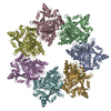 7t8cMC 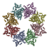 7t8bC M: atomic model generated by this map C: citing same article ( |
|---|---|
| Similar structure data |


-Links
| EMDB pages | EMDB (EBI/PDBe) / EMDataResource |
|---|---|
| Related items in Molecule of the Month |

-Map
| File | Download / File: emd_25744.map.gz / Format: CCP4 / Size: 244.1 MB / Type: IMAGE STORED AS FLOATING POINT NUMBER (4 BYTES) | ||||||||||||||||||||||||||||||||||||
|---|---|---|---|---|---|---|---|---|---|---|---|---|---|---|---|---|---|---|---|---|---|---|---|---|---|---|---|---|---|---|---|---|---|---|---|---|---|
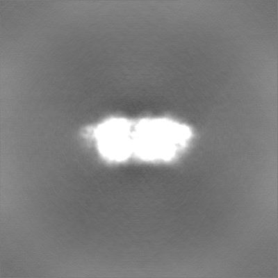
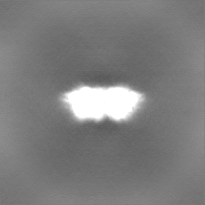
| Projections & slices | Image control
Images are generated by Spider. | ||||||||||||||||||||||||||||||||||||
| Voxel size | X=Y=Z: 1.058 Å | ||||||||||||||||||||||||||||||||||||
| Density |
| ||||||||||||||||||||||||||||||||||||
| Symmetry | Space group: 1 | ||||||||||||||||||||||||||||||||||||
| Details | EMDB XML:
|
 Z (Sec.)
 Y (Row.)
 X (Col.)


-Supplemental data
-Mask #1
| File | emd_25744_msk_1.map | ||||||||||||
|---|---|---|---|---|---|---|---|---|---|---|---|---|---|
| Projections & Slices |
| ||||||||||||
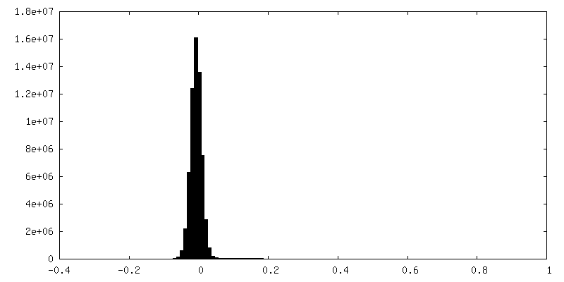
| Density Histograms |


-Half map: #2
| File | emd_25744_half_map_1.map | ||||||||||||
|---|---|---|---|---|---|---|---|---|---|---|---|---|---|
| Projections & Slices |
| ||||||||||||
| Density Histograms |


-Half map: #1
| File | emd_25744_half_map_2.map | ||||||||||||
|---|---|---|---|---|---|---|---|---|---|---|---|---|---|
| Projections & Slices |
| ||||||||||||
| Density Histograms |


- Sample components
-Entire : Heptameric Human Twinkle Clinical Variant W315L
| Entire | Name: Heptameric Human Twinkle Clinical Variant W315L |
|---|---|
| Components |
|
-Supramolecule #1: Heptameric Human Twinkle Clinical Variant W315L
| Supramolecule | Name: Heptameric Human Twinkle Clinical Variant W315L / type: complex / ID: 1 / Parent: 0 / Macromolecule list: all |
|---|---|
| Source (natural) | Organism: Homo sapiens (human) |
| Molecular weight | Theoretical: 504 KDa |
-Macromolecule #1: Twinkle mtDNA helicase
| Macromolecule | Name: Twinkle mtDNA helicase / type: protein_or_peptide / ID: 1 / Number of copies: 7 / Enantiomer: LEVO / EC number: DNA helicase |
|---|---|
| Source (natural) | Organism: Homo sapiens (human) |
| Molecular weight | Theoretical: 78.469422 KDa |
| Recombinant expression | Organism:  Escherichia coli (E. coli) Escherichia coli (E. coli) |
| Sequence | String: MWVLLRSGYP LRILLPLRGE WMGRRGLPRN LAPGPPRRRY RKETLQALDM PVLPVTATEI RQYLRGHGIP FQDGHSCLRA LSPFAESSQ LKGQTGVTTS FSLFIDKTTG HFLCMTSLAE GSWEDFQASV EGRGDGAREG FLLSKAPEFE DSEEVRRIWN R AIPLWELP ...String: MWVLLRSGYP LRILLPLRGE WMGRRGLPRN LAPGPPRRRY RKETLQALDM PVLPVTATEI RQYLRGHGIP FQDGHSCLRA LSPFAESSQ LKGQTGVTTS FSLFIDKTTG HFLCMTSLAE GSWEDFQASV EGRGDGAREG FLLSKAPEFE DSEEVRRIWN R AIPLWELP DQEEVQLADT MFGLTKVTDD TLKRFSVRYL RPARSLVFPW FSPGGSGLRG LKLLEAKCQG DGVSYEETTI PR PSAYHNL FGLPLISRRD AEVVLTSREL DSLALNQSTG LPTLTLPRGT TCLPPALLPY LEQFRRIVFW LGDDLRSLEA AKL FARKLN PKRCFLVRPG DQQPRPLEAL NGGFNLSRIL RTALPAWHKS IVSFRQLREE VLGELSNVEQ AAGLRWSRFP DLNR ILKGH RKGELTVFTG PTGSGKTTFI SEYALDLCSQ GVNTLWGSFE ISNVRLARVM LTQFAEGRLE DQLDKYDHWA DRFED LPLY FMTFHGQQSI RTVIDTMQHA VYVYDICHVI IDNLQFMMGH EQLSTDRIAA QDYIIGVFRK FATDNNCHVT LVIHPR KED DDKELQTASI FGSAKASQEA DNVLILQDRK LVTGPGKRYL QVSKNRFDGD VGVFPLEFNK NSLTFSIPPK NKARLKK IK DDTGPVAKKP SSGKKGATTQ NSEICSGQAP TPDQPDTSKR SKAAALEHHH HHH UniProtKB: Twinkle mtDNA helicase |
-Experimental details
-Structure determination
| Method | cryo EM |
|---|---|
 Processing Processing | single particle reconstruction |
| Aggregation state | particle |
-Sample preparation
| Concentration | 1.2 mg/mL |
|---|---|
| Buffer | pH: 8 |
| Vitrification | Cryogen name: ETHANE |
- Electron microscopy
| Microscope | TFS KRIOS |
|---|---|
| Electron beam | Acceleration voltage: 300 kV / Electron source: FIELD EMISSION GUN |
| Electron optics | Illumination mode: FLOOD BEAM / Imaging mode: BRIGHT FIELDBright-field microscopy / Nominal defocus max: 2.5 µm / Nominal defocus min: 1.0 µm |
| Image recording | Film or detector model: GATAN K3 BIOQUANTUM (6k x 4k) / Average electron dose: 60.0 e/Å2 |
| Experimental equipment |  Model: Titan Krios / Image courtesy: FEI Company |
-Image processing
| Startup model | Type of model: OTHER |
|---|---|
| Initial angle assignment | Type: MAXIMUM LIKELIHOOD |
| Final angle assignment | Type: MAXIMUM LIKELIHOOD |
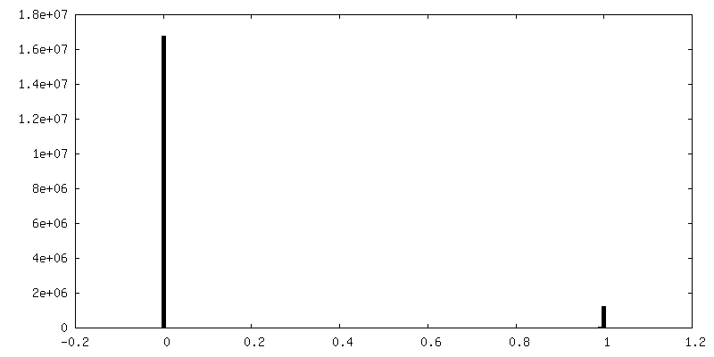
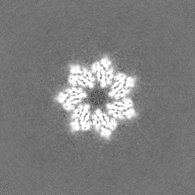
| Final reconstruction | Applied symmetry - Point group: C7 (7 fold cyclic) / Resolution.type: BY AUTHOR / Resolution: 4.5 Å / Resolution method: FSC 0.143 CUT-OFF / Number images used: 55655 |
| FSC plot (resolution estimation) |  |
