 Movie
Movie Controller


+ Open data

- Basic information
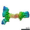
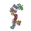
| Entry | Database: EMDB / ID: EMD-25128 | ||||||||||||
|---|---|---|---|---|---|---|---|---|---|---|---|---|---|
| Title | Structure of Xenopus laevis CRL2Lrr1 (State 2) | ||||||||||||
 Map data Map data | |||||||||||||
 Sample Sample |
| ||||||||||||
 Keywords Keywords | Cullin RING E3 ubiquitin ligase / DNA replication termination /  LIGASE LIGASE | ||||||||||||
| Function / homology |  Function and homology information Function and homology informationcullin-RING ubiquitin ligase complex / elongin complex / VCB complex / transcription elongation by RNA polymerase II / protein-macromolecule adaptor activity / ubiquitin-dependent protein catabolic process / protein ubiquitination / ubiquitin protein ligase binding / zinc ion bindingSimilarity search - Function | ||||||||||||
| Biological species | Xenopus laevis (African clawed frog) | ||||||||||||
| Method | single particle reconstruction / cryo EM / Resolution: 3.5 Å | ||||||||||||
 Authors Authors | Zhou H / Brown A | ||||||||||||
| Funding support |  United States, 3 items United States, 3 items
| ||||||||||||
 Citation Citation | Journal: Nucleic Acids Res / Year: 2021 Title: Structure of CRL2Lrr1, the E3 ubiquitin ligase that promotes DNA replication termination in vertebrates. Authors: Haixia Zhou / Manal S Zaher / Johannes C Walter / Alan Brown / Abstract: When vertebrate replisomes from neighboring origins converge, the Mcm7 subunit of the replicative helicase, CMG, is ubiquitylated by the E3 ubiquitin ligase, CRL2Lrr1. Polyubiquitylated CMG is then ...When vertebrate replisomes from neighboring origins converge, the Mcm7 subunit of the replicative helicase, CMG, is ubiquitylated by the E3 ubiquitin ligase, CRL2Lrr1. Polyubiquitylated CMG is then disassembled by the p97 ATPase, leading to replication termination. To avoid premature replisome disassembly, CRL2Lrr1 is only recruited to CMGs after they converge, but the underlying mechanism is unclear. Here, we use cryogenic electron microscopy to determine structures of recombinant Xenopus laevis CRL2Lrr1 with and without neddylation. The structures reveal that CRL2Lrr1 adopts an unusually open architecture, in which the putative substrate-recognition subunit, Lrr1, is located far from the catalytic module that catalyzes ubiquitin transfer. We further demonstrate that a predicted, flexible pleckstrin homology domain at the N-terminus of Lrr1 is essential to target CRL2Lrr1 to terminated CMGs. We propose a hypothetical model that explains how CRL2Lrr1's catalytic module is positioned next to the ubiquitylation site on Mcm7, and why CRL2Lrr1 binds CMG only after replisomes converge. | ||||||||||||
| History |
|
- Structure visualization
| Movie |
Movie viewer |
|---|---|
| Structure viewer | EM map: SurfViewMolmilJmol/JSmol |
| Supplemental images |

- Downloads & links
-EMDB archive
| Map data | emd_25128.map.gz | 4 MB | EMDB map data format | |
|---|---|---|---|---|
| Header (meta data) | emd-25128-v30.xmlemd-25128.xml | 17.3 KB 17.3 KB | Display Display | EMDB header |
| FSC (resolution estimation) | emd_25128_fsc.xml | 11.4 KB | Display | FSC data file |
| Images |  emd_25128.png emd_25128.png | 68.4 KB | ||
| Masks | emd_25128_msk_1.map | 125 MB | Mask map | |
| Filedesc metadata | emd-25128.cif.gz | 6.7 KB | ||
| Archive directory |  http://ftp.pdbj.org/pub/emdb/structures/EMD-25128ftp://ftp.pdbj.org/pub/emdb/structures/EMD-25128 http://ftp.pdbj.org/pub/emdb/structures/EMD-25128ftp://ftp.pdbj.org/pub/emdb/structures/EMD-25128 | HTTPS FTP |
-Related structure data
| Related structure data |  7shlMC  7shkC M: atomic model generated by this map C: citing same article ( |
|---|---|
| Similar structure data |


-Links
| EMDB pages | EMDB (EBI/PDBe) / EMDataResource |
|---|---|
| Related items in Molecule of the Month |


-Map
| File | Download / File: emd_25128.map.gz / Format: CCP4 / Size: 125 MB / Type: IMAGE STORED AS FLOATING POINT NUMBER (4 BYTES) | ||||||||||||||||||||||||||||||||||||||||||||||||||||||||||||||||||||
|---|---|---|---|---|---|---|---|---|---|---|---|---|---|---|---|---|---|---|---|---|---|---|---|---|---|---|---|---|---|---|---|---|---|---|---|---|---|---|---|---|---|---|---|---|---|---|---|---|---|---|---|---|---|---|---|---|---|---|---|---|---|---|---|---|---|---|---|---|---|
| Voxel size | X=Y=Z: 0.825 Å | ||||||||||||||||||||||||||||||||||||||||||||||||||||||||||||||||||||
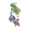
| Density |
| ||||||||||||||||||||||||||||||||||||||||||||||||||||||||||||||||||||
| Symmetry | Space group: 1 | ||||||||||||||||||||||||||||||||||||||||||||||||||||||||||||||||||||
| Details | EMDB XML:
CCP4 map header:
| ||||||||||||||||||||||||||||||||||||||||||||||||||||||||||||||||||||
-Supplemental data
-Mask #1
| File | emd_25128_msk_1.map | ||||||||||||
|---|---|---|---|---|---|---|---|---|---|---|---|---|---|
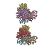
| Projections & Slices |
| ||||||||||||
| Density Histograms |
 Z
Z Y
Y X
X


- Sample components
-Entire : CRL2Lrr1
| Entire | Name: CRL2Lrr1 |
|---|---|
| Components |
|
-Supramolecule #1: CRL2Lrr1
| Supramolecule | Name: CRL2Lrr1 / type: complex / ID: 1 / Parent: 0 / Macromolecule list: #1-#5 |
|---|---|
| Source (natural) | Organism: Xenopus laevis (African clawed frog) |
-Macromolecule #1: CULLIN_2 domain-containing protein
| Macromolecule | Name: CULLIN_2 domain-containing protein / type: protein_or_peptide / ID: 1 / Number of copies: 1 / Enantiomer: LEVO |
|---|---|
| Source (natural) | Organism: Xenopus laevis (African clawed frog) |
| Molecular weight | Theoretical: 87.244117 KDa |
| Recombinant expression | Organism:   Spodoptera frugiperda (fall armyworm) Spodoptera frugiperda (fall armyworm) |
| Sequence | String: MSLKPRVVDF DETWNKLLTT IKAVVMLDYV ERATWNDRFS DIYALCVAYP EPLGERLYTE TKIFLENHVQ QLHTRVLDSA EQVLVMYFR YWEEYSRGAD YMDCLYRYLN TQYIKKNKLT EADLQYGYGG VDMNEPLMEI GELALDLWRK LMIEPLQDTL L IMLLREIK ...String: MSLKPRVVDF DETWNKLLTT IKAVVMLDYV ERATWNDRFS DIYALCVAYP EPLGERLYTE TKIFLENHVQ QLHTRVLDSA EQVLVMYFR YWEEYSRGAD YMDCLYRYLN TQYIKKNKLT EADLQYGYGG VDMNEPLMEI GELALDLWRK LMIEPLQDTL L IMLLREIK RDRCGEDPNQ KVIHGVINSF VHVEQYKKKF PLKFYQEIFE SPFLAETGEY YKQEASNLLQ ESNCSQYMEK IL GRLKDEE IRCRKYLHPS SYNKVIHECQ QRMVADHLQF LHAECHNIIR QERRNDMANM YTLLRAVSSG LPHMIQELQN HIH DEGLRA ISNLSQENMP TQFVESVLEV HSKFVQLVNC VLNGDQHFMS ALDKALTCVV NYREPKSVCK APELLAKYCD NMLK KSAKG MTENEVEDKL TSFITVFKYI DDKDVFQKFY ARMLAKRLIH GLSMSMDSEE TMINKLKQAC GYEFTSKLHR MYTDM SVSA DLNNKFNNFI KSQDTVIDLG ISFQIYVLQA GAWPLTQAPS STFAIPQELE KSVQMFELFY NQHFSGRKLT WLHYLC TGE VKMNYLCKPY VAMVTTYQMA VLLAFNNSEI ITYKELQDST QMNEKELTKT IKSLLDVKMI NHDSDKEDIE GESTFSL NM NFSSKRTKFK ITTPMQKDTP QEVEQTRSAV DEDRKMYLQA AIVRIMKARK VLRHNALIQE VISQSRARFN PSISMIKK C IEVLIDKQYI ERSQASADEY SYVA UniProtKB: CULLIN_2 domain-containing protein |
-Macromolecule #2: Tceb2-prov protein
| Macromolecule | Name: Tceb2-prov protein / type: protein_or_peptide / ID: 2 / Number of copies: 1 / Enantiomer: LEVO |
|---|---|
| Source (natural) | Organism: Xenopus laevis (African clawed frog) |
| Molecular weight | Theoretical: 13.325927 KDa |
| Recombinant expression | Organism: Spodoptera frugiperda (fall armyworm) |
| Sequence | String: MDVFLMIRHH KTTIFTDAKE NTTVYELKRI VEGILKRPPE DQKLYKDDQL LDDNKTLGDC GFTSQTARPQ APATVGLAFR SSGDSFEPL RVEPFSSPPE LPDVMKPQET SGSANEQAVQ UniProtKB: Elongin B L homeolog |
-Macromolecule #3: Elongin-C
| Macromolecule | Name: Elongin-C / type: protein_or_peptide / ID: 3 / Number of copies: 1 / Enantiomer: LEVO |
|---|---|
| Source (natural) | Organism: Xenopus laevis (African clawed frog) |
| Molecular weight | Theoretical: 12.485135 KDa |
| Recombinant expression | Organism: Spodoptera frugiperda (fall armyworm) |
| Sequence | String: MDGEEKTYGG CEGPDAMYVK LISSDGHEFI VKREHALTSG TIKAMLSGPG QFAENETNEV NFREIPSHVL SKVCMYFTYK VRYTNSSTE IPEFPIAPEI ALELLMAANF LDC UniProtKB: Elongin-C |
-Macromolecule #4: Lrr1
| Macromolecule | Name: Lrr1 / type: protein_or_peptide / ID: 4 / Number of copies: 1 / Enantiomer: LEVO |
|---|---|
| Source (natural) | Organism: Xenopus laevis (African clawed frog) |
| Molecular weight | Theoretical: 46.882258 KDa |
| Recombinant expression | Organism: Spodoptera frugiperda (fall armyworm) |
| Sequence | String: MKLQCEVEVI NRMLPTFGLK NRGKGTRAVL SVGRQEGKRG AAYLMICTLK DKSGSRYKLE NNIEQLFTRF VGEGKATLRL KEPALDICL SKAEICGLRN FISTVGLANK GTDIGTVSLP RLTPAKTSEI EKPRSKLFIT TKKDYPITKS FPYSLEHLQV S YCKLARVD ...String: MKLQCEVEVI NRMLPTFGLK NRGKGTRAVL SVGRQEGKRG AAYLMICTLK DKSGSRYKLE NNIEQLFTRF VGEGKATLRL KEPALDICL SKAEICGLRN FISTVGLANK GTDIGTVSLP RLTPAKTSEI EKPRSKLFIT TKKDYPITKS FPYSLEHLQV S YCKLARVD MRMLCLKKLQ KLDLSNNHIK KLPKTIGDLV CLQELILNHN FLESFEVVLC STTLRDTLKS LDLSANKLKA LP VQICNFK ELVSLKLDEN ELLQLPFPIG QLSKLRFLSA TKNNLQCLPN TFKKLTLENL DLFGNPFMQA TPLVPDIQLK IPL PLLETA ARATLKYRIP YGPHLIPATL CQDLSLAKTC DCGLPCLNSF IQTIVLMNLH QVSQTVVLVD TMGGTDGPIV CYFC SLTCY SQFLDKYLQS TRV UniProtKB: Leucine rich repeat protein 1 L homeolog isoform X1 |
-Macromolecule #5: RING-type domain-containing protein
| Macromolecule | Name: RING-type domain-containing protein / type: protein_or_peptide / ID: 5 / Number of copies: 1 / Enantiomer: LEVO |
|---|---|
| Source (natural) | Organism: Xenopus laevis (African clawed frog) |
| Molecular weight | Theoretical: 12.277985 KDa |
| Recombinant expression | Organism: Spodoptera frugiperda (fall armyworm) |
| Sequence | String: MAAAMDVDTP SGANNSAGKK RFEVKKWNAV ALWAWDIVVD NCAICRNHIM DLCIECQANQ ASATSEECTV AWGVCNHAFH FHCISRWLK TRQVCPLDNR EWEFQKYVG UniProtKB: RING-type domain-containing protein |
-Macromolecule #6: ZINC ION
| Macromolecule | Name: ZINC ION / type: ligand / ID: 6 / Number of copies: 1 / Formula: ZN |
|---|---|
| Molecular weight | Theoretical: 65.409 Da |
-Experimental details
-Structure determination
| Method | cryo EM |
|---|---|
 Processing Processing | single particle reconstruction |
| Aggregation state | particle |
-Sample preparation
| Concentration | 0.8 mg/mL | ||||||||||||
|---|---|---|---|---|---|---|---|---|---|---|---|---|---|
| Buffer | pH: 7.6 Component:
Details: TCEP is freshly added. | ||||||||||||
| Grid | Model: C-flat-1.2/1.3 / Material: COPPER / Mesh: 400 / Pretreatment - Type: GLOW DISCHARGE | ||||||||||||
| Vitrification | Cryogen name: NITROGEN / Chamber humidity: 100 % / Chamber temperature: 281.2 K / Instrument: FEI VITROBOT MARK IV / Details: Blot 6 seconds with the force 12. |
- Electron microscopy
| Microscope | FEI TITAN KRIOS |
|---|---|
| Electron beam | Acceleration voltage: 300 kV / Electron source: FIELD EMISSION GUN |
| Electron optics | Illumination mode: FLOOD BEAM / Imaging mode: BRIGHT FIELDBright-field microscopy |
| Sample stage | Specimen holder model: FEI TITAN KRIOS AUTOGRID HOLDER |
| Image recording | Film or detector model: GATAN K3 BIOQUANTUM (6k x 4k) / Average electron dose: 54.5 e/Å2 |
| Experimental equipment |  Model: Titan Krios / Image courtesy: FEI Company |
-Image processing
| Startup model | Type of model: OTHER / Details: Ab initio model |
|---|---|
| Initial angle assignment | Type: ANGULAR RECONSTITUTION |
| Final angle assignment | Type: ANGULAR RECONSTITUTION |
| Final reconstruction | Resolution.type: BY AUTHOR / Resolution: 3.5 Å / Resolution method: FSC 0.143 CUT-OFF / Software - Name: RELION (ver. 3.1) / Number images used: 406755 |
| FSC plot (resolution estimation) |  |
-Atomic model buiding 1
| Refinement | Space: REAL / Protocol: OTHER / Target criteria: Correlation coefficient |
|---|---|
| Output model | PDB-7shl: |
