National Institutes of Health/National Institute Of Allergy and Infectious Diseases (NIH/NIAID)
P01-AI138938-S1
United States
Citation
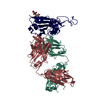
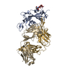
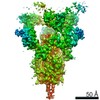
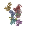
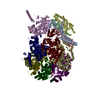
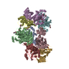
Journal: Cell Rep / Year: 2021 Title: Broad cross-reactivity across sarbecoviruses exhibited by a subset of COVID-19 donor-derived neutralizing antibodies. Authors: Claudia A Jette / Alexander A Cohen / Priyanthi N P Gnanapragasam / Frauke Muecksch / Yu E Lee / Kathryn E Huey-Tubman / Fabian Schmidt / Theodora Hatziioannou / Paul D Bieniasz / Michel C ...Authors: Claudia A Jette / Alexander A Cohen / Priyanthi N P Gnanapragasam / Frauke Muecksch / Yu E Lee / Kathryn E Huey-Tubman / Fabian Schmidt / Theodora Hatziioannou / Paul D Bieniasz / Michel C Nussenzweig / Anthony P West / Jennifer R Keeffe / Pamela J Bjorkman / Christopher O Barnes / Abstract: Many anti-severe acute respiratory syndrome coronavirus 2 (anti-SARS-CoV-2) neutralizing antibodies target the angiotensin-converting enzyme 2 (ACE2) binding site on viral spike receptor-binding ...Many anti-severe acute respiratory syndrome coronavirus 2 (anti-SARS-CoV-2) neutralizing antibodies target the angiotensin-converting enzyme 2 (ACE2) binding site on viral spike receptor-binding domains (RBDs). Potent antibodies recognize exposed variable epitopes, often rendering them ineffective against other sarbecoviruses and SARS-CoV-2 variants. Class 4 anti-RBD antibodies against a less-exposed, but more-conserved, cryptic epitope could recognize newly emergent zoonotic sarbecoviruses and variants, but they usually show only weak neutralization potencies. Here, we characterize two class 4 anti-RBD antibodies derived from coronavirus disease 2019 (COVID-19) donors that exhibit breadth and potent neutralization of zoonotic coronaviruses and SARS-CoV-2 variants. C118-RBD and C022-RBD structures reveal orientations that extend from the cryptic epitope to occlude ACE2 binding and CDRH3-RBD main-chain H-bond interactions that extend an RBD β sheet, thus reducing sensitivity to RBD side-chain changes. A C118-spike trimer structure reveals rotated RBDs that allow access to the cryptic epitope and the potential for intra-spike crosslinking to increase avidity. These studies facilitate vaccine design and illustrate potential advantages of class 4 RBD-binding antibody therapeutics.
History
Deposition
Jul 22, 2021
-
Header (metadata) release
Sep 22, 2021
-
Map release
Sep 22, 2021
-
Update
Oct 13, 2021
-
Current status
Oct 13, 2021
Processing site: RCSB / Status: Released
-
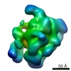
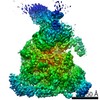
Structure visualization
Movie
Surface view with section colored by density value
Film or detector model: GATAN K3 (6k x 4k) / Number grids imaged: 1 / Number real images: 2970 / Average exposure time: 3.6 sec. / Average electron dose: 60.0 e/Å2
Experimental equipment
Model: Talos Arctica / Image courtesy: FEI Company
-
Image processing
Startup model
Type of model: OTHER / Details: ab initio
Initial angle assignment
Type: MAXIMUM LIKELIHOOD
Final 3D classification
Number classes: 4 / Software - Name: cryoSPARC (ver. 3.1)
Final angle assignment
Type: MAXIMUM LIKELIHOOD
Final reconstruction
Number classes used: 1 / Applied symmetry - Point group: C3 (3 fold cyclic) / Algorithm: FOURIER SPACE / Resolution.type: BY AUTHOR / Resolution: 3.45 Å / Resolution method: FSC 0.143 CUT-OFF / Software - Name: cryoSPARC (ver. 3.1) / Number images used: 53728
FSC plot (resolution estimation)
+
About Yorodumi
-
News
-
Feb 9, 2022. New format data for meta-information of EMDB entries
New format data for meta-information of EMDB entries
Version 3 of the EMDB header file is now the official format.
The previous official version 1.9 will be removed from the archive.
In the structure databanks used in Yorodumi, some data are registered as the other names, "COVID-19 virus" and "2019-nCoV". Here are the details of the virus and the list of structure data.
Jan 31, 2019. EMDB accession codes are about to change! (news from PDBe EMDB page)
EMDB accession codes are about to change! (news from PDBe EMDB page)
The allocation of 4 digits for EMDB accession codes will soon come to an end. Whilst these codes will remain in use, new EMDB accession codes will include an additional digit and will expand incrementally as the available range of codes is exhausted. The current 4-digit format prefixed with “EMD-” (i.e. EMD-XXXX) will advance to a 5-digit format (i.e. EMD-XXXXX), and so on. It is currently estimated that the 4-digit codes will be depleted around Spring 2019, at which point the 5-digit format will come into force.
The EM Navigator/Yorodumi systems omit the EMD- prefix.
Related info.:Q: What is EMD? / ID/Accession-code notation in Yorodumi/EM Navigator
Yorodumi is a browser for structure data from EMDB, PDB, SASBDB, etc.
This page is also the successor to EM Navigator detail page, and also detail information page/front-end page for Omokage search.
The word "yorodu" (or yorozu) is an old Japanese word meaning "ten thousand". "mi" (miru) is to see.
Related info.:EMDB / PDB / SASBDB / Comparison of 3 databanks / Yorodumi Search / Aug 31, 2016. New EM Navigator & Yorodumi / Yorodumi Papers / Jmol/JSmol / Function and homology information / Changes in new EM Navigator and Yorodumi
 Movie
Movie Controller

 Yorodumi
 Open data
 Basic information
 Map data
 Sample
 Function and homology information
 membrane fusion / positive regulation of viral entry into host cell / receptor-mediated virion attachment to host cell /


 Authors
 United States, 1 items
  Citation
 Structure visualization
 Downloads & links
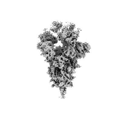 emd_24504.png
 http://ftp.pdbj.org/pub/emdb/structures/EMD-24504
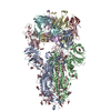


 Z
Z Y
Y X
X


 Sample components
 Processing
 Electron microscopy

