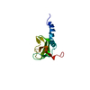
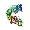
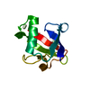
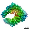
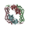
Journal: J Cell Sci / Year: 2015 Title: The structure of the complex between α-tubulin, TBCE and TBCB reveals a tubulin dimer dissociation mechanism. Authors: Marina Serna / Gerardo Carranza / Jaime Martín-Benito / Robert Janowski / Albert Canals / Miquel Coll / Juan Carlos Zabala / José María Valpuesta / Abstract: Tubulin proteostasis is regulated by a group of molecular chaperones termed tubulin cofactors (TBC). Whereas tubulin heterodimer formation is well-characterized biochemically, its dissociation ...Tubulin proteostasis is regulated by a group of molecular chaperones termed tubulin cofactors (TBC). Whereas tubulin heterodimer formation is well-characterized biochemically, its dissociation pathway is not clearly understood. Here, we carried out biochemical assays to dissect the role of the human TBCE and TBCB chaperones in α-tubulin-β-tubulin dissociation. We used electron microscopy and image processing to determine the three-dimensional structure of the human TBCE, TBCB and α-tubulin (αEB) complex, which is formed upon α-tubulin-β-tubulin heterodimer dissociation by the two chaperones. Docking the atomic structures of domains of these proteins, including the TBCE UBL domain, as we determined by X-ray crystallography, allowed description of the molecular architecture of the αEB complex. We found that heterodimer dissociation is an energy-independent process that takes place through a disruption of the α-tubulin-β-tubulin interface that is caused by a steric interaction between β-tubulin and the TBCE cytoskeleton-associated protein glycine-rich (CAP-Gly) and leucine-rich repeat (LRR) domains. The protruding arrangement of chaperone ubiquitin-like (UBL) domains in the αEB complex suggests that there is a direct interaction of this complex with the proteasome, thus mediating α-tubulin degradation.
History
Deposition
Aug 27, 2013
-
Header (metadata) release
Sep 18, 2013
-
Map release
Sep 10, 2014
-
Update
Aug 26, 2015
-
Current status
Aug 26, 2015
Processing site: PDBe / Status: Released
-
Structure visualization
Movie
Surface view with section colored by density value
Legacy - Astigmatism: Objective lens astigmatism was corrected at 100,000 times magnification
Date
Nov 1, 2010
Image recording
Category: FILM / Film or detector model: KODAK SO-163 FILM / Digitization - Scanner: ZEISS SCAI / Digitization - Sampling interval: 14 µm / Number real images: 85 / Average electron dose: 10 e/Å2
-
Image processing
CTF correction
Details: N. Grigorieff CTFFIND3
Final reconstruction
Applied symmetry - Point group: C1 (asymmetric) / Algorithm: OTHER / Resolution.type: BY AUTHOR / Resolution: 21.0 Å / Resolution method: FSC 0.33 CUT-OFF / Software - Name: EMAN, Spider, XMIPP Details: Individual particles were manually selected using XMIPP software package. Number images used: 26129
Details
The particles were manually selected using the XMIPP program
In the structure databanks used in Yorodumi, some data are registered as the other names, "COVID-19 virus" and "2019-nCoV". Here are the details of the virus and the list of structure data.
Jan 31, 2019. EMDB accession codes are about to change! (news from PDBe EMDB page)
EMDB accession codes are about to change! (news from PDBe EMDB page)
The allocation of 4 digits for EMDB accession codes will soon come to an end. Whilst these codes will remain in use, new EMDB accession codes will include an additional digit and will expand incrementally as the available range of codes is exhausted. The current 4-digit format prefixed with “EMD-” (i.e. EMD-XXXX) will advance to a 5-digit format (i.e. EMD-XXXXX), and so on. It is currently estimated that the 4-digit codes will be depleted around Spring 2019, at which point the 5-digit format will come into force.
The EM Navigator/Yorodumi systems omit the EMD- prefix.
Related info.:Q: What is EMD? / ID/Accession-code notation in Yorodumi/EM Navigator
Yorodumi is a browser for structure data from EMDB, PDB, SASBDB, etc.
This page is also the successor to EM Navigator detail page, and also detail information page/front-end page for Omokage search.
The word "yorodu" (or yorozu) is an old Japanese word meaning "ten thousand". "mi" (miru) is to see.
Related info.:EMDB / PDB / SASBDB / Comparison of 3 databanks / Yorodumi Search / Aug 31, 2016. New EM Navigator & Yorodumi / Yorodumi Papers / Jmol/JSmol / Function and homology information / Changes in new EM Navigator and Yorodumi
 Movie
Movie Controller

 Yorodumi
 Open data
 Basic information
 Map data
 Sample
 Keywords
 protein folding /
  Function and homology information

 Authors
 Citation
 Structure visualization
 Downloads & links
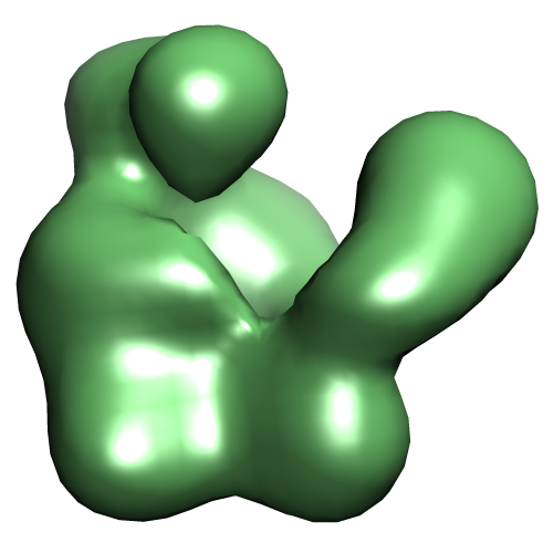 EMD-2447.png
 http://ftp.pdbj.org/pub/emdb/structures/EMD-2447
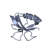


 Sample components


 Processing
 Electron microscopy