 Movie
Movie Controller

[English] 日本語
 Yorodumi
- EMDB-23812: Structure of the apo phosphoinositide 3-kinase p110 gamma (PIK3CG... -
+ Open data

- Basic information
| Entry | Database: EMDB / ID: EMD-23812 | |||||||||
|---|---|---|---|---|---|---|---|---|---|---|
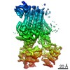
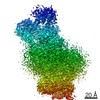
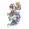
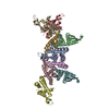
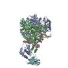
| Title | Structure of the apo phosphoinositide 3-kinase p110 gamma (PIK3CG) p101 (PIK3R5) complex | |||||||||
 Map data Map data | ||||||||||
 Sample Sample |
| |||||||||
| Function / homology |  Function and homology information Function and homology information: / secretory granule localization / negative regulation of triglyceride catabolic process / natural killer cell chemotaxis / neutrophil extravasation /  phosphatidylinositol-4-phosphate 3-kinase / regulation of calcium ion transmembrane transport / positive regulation of acute inflammatory response / respiratory burst involved in defense response / negative regulation of cardiac muscle contraction ...: / secretory granule localization / negative regulation of triglyceride catabolic process / natural killer cell chemotaxis / neutrophil extravasation / phosphatidylinositol-4-phosphate 3-kinase / regulation of calcium ion transmembrane transport / positive regulation of acute inflammatory response / respiratory burst involved in defense response / negative regulation of cardiac muscle contraction / 1-phosphatidylinositol-3-kinase regulator activity / T cell chemotaxis / negative regulation of fibroblast apoptotic process / phosphatidylinositol 3-kinase complex, class IB / sphingosine-1-phosphate receptor signaling pathway / phosphatidylinositol 3-kinase complex / dendritic cell chemotaxis / 1-phosphatidylinositol-4-phosphate 3-kinase activity / 1-phosphatidylinositol-4,5-bisphosphate 3-kinase activity / phosphatidylinositol-4,5-bisphosphate 3-kinase / phosphatidylinositol 3-kinase / phosphatidylinositol 3-kinase complex, class IA / phosphatidylinositol-3-phosphate biosynthetic process / 1-phosphatidylinositol-3-kinase activity / mast cell degranulation / Erythropoietin activates Phosphoinositide-3-kinase (PI3K) / hepatocyte apoptotic process / positive regulation of Rac protein signal transduction / regulation of cell adhesion mediated by integrin / Synthesis of PIPs at the plasma membrane / phosphatidylinositol phosphate biosynthetic process / centriolar satellite / regulation of angiogenesis / T cell proliferation / cellular response to cAMP / GPVI-mediated activation cascade / T cell activation / ephrin receptor binding / neutrophil chemotaxis / positive regulation of endothelial cell migration / phosphatidylinositol 3-kinase/protein kinase B signal transduction / positive regulation of cytokine production / G-protein beta/gamma-subunit complex binding / positive regulation of MAP kinase activity / platelet aggregation / endocytosis / G beta:gamma signalling through PI3Kgamma / Signaling by CSF1 (M-CSF) in myeloid cells / kinase activity / positive regulation of cytosolic calcium ion concentration / angiogenesis / adaptive immune response / positive regulation of phosphatidylinositol 3-kinase/protein kinase B signal transduction / non-specific serine/threonine protein kinase / protein kinase activity / immune response / inflammatory response / G protein-coupled receptor signaling pathway / phosphorylation / protein serine kinase activity / innate immune response / protein serine/threonine kinase activity / ATP binding / membrane / identical protein binding / nucleus / plasma membrane / cytosol / cytoplasm phosphatidylinositol-4-phosphate 3-kinase / regulation of calcium ion transmembrane transport / positive regulation of acute inflammatory response / respiratory burst involved in defense response / negative regulation of cardiac muscle contraction ...: / secretory granule localization / negative regulation of triglyceride catabolic process / natural killer cell chemotaxis / neutrophil extravasation / phosphatidylinositol-4-phosphate 3-kinase / regulation of calcium ion transmembrane transport / positive regulation of acute inflammatory response / respiratory burst involved in defense response / negative regulation of cardiac muscle contraction / 1-phosphatidylinositol-3-kinase regulator activity / T cell chemotaxis / negative regulation of fibroblast apoptotic process / phosphatidylinositol 3-kinase complex, class IB / sphingosine-1-phosphate receptor signaling pathway / phosphatidylinositol 3-kinase complex / dendritic cell chemotaxis / 1-phosphatidylinositol-4-phosphate 3-kinase activity / 1-phosphatidylinositol-4,5-bisphosphate 3-kinase activity / phosphatidylinositol-4,5-bisphosphate 3-kinase / phosphatidylinositol 3-kinase / phosphatidylinositol 3-kinase complex, class IA / phosphatidylinositol-3-phosphate biosynthetic process / 1-phosphatidylinositol-3-kinase activity / mast cell degranulation / Erythropoietin activates Phosphoinositide-3-kinase (PI3K) / hepatocyte apoptotic process / positive regulation of Rac protein signal transduction / regulation of cell adhesion mediated by integrin / Synthesis of PIPs at the plasma membrane / phosphatidylinositol phosphate biosynthetic process / centriolar satellite / regulation of angiogenesis / T cell proliferation / cellular response to cAMP / GPVI-mediated activation cascade / T cell activation / ephrin receptor binding / neutrophil chemotaxis / positive regulation of endothelial cell migration / phosphatidylinositol 3-kinase/protein kinase B signal transduction / positive regulation of cytokine production / G-protein beta/gamma-subunit complex binding / positive regulation of MAP kinase activity / platelet aggregation / endocytosis / G beta:gamma signalling through PI3Kgamma / Signaling by CSF1 (M-CSF) in myeloid cells / kinase activity / positive regulation of cytosolic calcium ion concentration / angiogenesis / adaptive immune response / positive regulation of phosphatidylinositol 3-kinase/protein kinase B signal transduction / non-specific serine/threonine protein kinase / protein kinase activity / immune response / inflammatory response / G protein-coupled receptor signaling pathway / phosphorylation / protein serine kinase activity / innate immune response / protein serine/threonine kinase activity / ATP binding / membrane / identical protein binding / nucleus / plasma membrane / cytosol / cytoplasmSimilarity search - Function | |||||||||
| Biological species |  Homo sapiens (human) Homo sapiens (human) | |||||||||
| Method | single particle reconstruction / cryo EM / Resolution: 3.36 Å | |||||||||
 Authors Authors | Burke JE / Dalwadi U / Rathinaswamy MK / Yip CK | |||||||||
| Funding support |  Canada, 1 items Canada, 1 items
| |||||||||
 Citation Citation | Journal: Sci Adv / Year: 2021 Title: Structure of the phosphoinositide 3-kinase (PI3K) p110γ-p101 complex reveals molecular mechanism of GPCR activation. Authors: Manoj K Rathinaswamy / Udit Dalwadi / Kaelin D Fleming / Carson Adams / Jordan T B Stariha / Els Pardon / Minkyung Baek / Oscar Vadas / Frank DiMaio / Jan Steyaert / Scott D Hansen / Calvin ...Authors: Manoj K Rathinaswamy / Udit Dalwadi / Kaelin D Fleming / Carson Adams / Jordan T B Stariha / Els Pardon / Minkyung Baek / Oscar Vadas / Frank DiMaio / Jan Steyaert / Scott D Hansen / Calvin K Yip / John E Burke /    Abstract: The class IB phosphoinositide 3-kinase (PI3K), PI3Kγ, is a master regulator of immune cell function and a promising drug target for both cancer and inflammatory diseases. Critical to PI3Kγ function ...The class IB phosphoinositide 3-kinase (PI3K), PI3Kγ, is a master regulator of immune cell function and a promising drug target for both cancer and inflammatory diseases. Critical to PI3Kγ function is the association of the p110γ catalytic subunit to either a p101 or p84 regulatory subunit, which mediates activation by G protein-coupled receptors. Here, we report the cryo-electron microscopy structure of a heterodimeric PI3Kγ complex, p110γ-p101. This structure reveals a unique assembly of catalytic and regulatory subunits that is distinct from other class I PI3K complexes. p101 mediates activation through its Gβγ-binding domain, recruiting the heterodimer to the membrane and allowing for engagement of a secondary Gβγ-binding site in p110γ. Mutations at the p110γ-p101 and p110γ-adaptor binding domain interfaces enhanced Gβγ activation. A nanobody that specifically binds to the p101-Gβγ interface blocks activation, providing a novel tool to study and target p110γ-p101-specific signaling events in vivo. | |||||||||
| History |
|
- Structure visualization
| Movie |
Movie viewer |
|---|---|
| Structure viewer | EM map: SurfViewMolmilJmol/JSmol |
| Supplemental images |

- Downloads & links
-EMDB archive
| Map data | emd_23812.map.gz | 97.3 MB | EMDB map data format | |
|---|---|---|---|---|
| Header (meta data) | emd-23812-v30.xmlemd-23812.xml | 14.6 KB 14.6 KB | Display Display | EMDB header |
| FSC (resolution estimation) | emd_23812_fsc.xml | 10.5 KB | Display | FSC data file |
| Images | 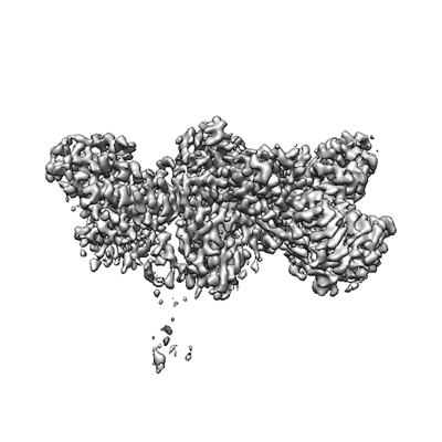 emd_23812.png emd_23812.png | 97.9 KB | ||
| Archive directory |  http://ftp.pdbj.org/pub/emdb/structures/EMD-23812ftp://ftp.pdbj.org/pub/emdb/structures/EMD-23812 http://ftp.pdbj.org/pub/emdb/structures/EMD-23812ftp://ftp.pdbj.org/pub/emdb/structures/EMD-23812 | HTTPS FTP |
-Related structure data
| Related structure data | |
|---|---|
| Similar structure data |


-Links
| EMDB pages | EMDB (EBI/PDBe) / EMDataResource |
|---|---|
| Related items in Molecule of the Month |


-Map
| File | Download / File: emd_23812.map.gz / Format: CCP4 / Size: 103 MB / Type: IMAGE STORED AS FLOATING POINT NUMBER (4 BYTES) | ||||||||||||||||||||||||||||||||||||||||||||||||||||||||||||
|---|---|---|---|---|---|---|---|---|---|---|---|---|---|---|---|---|---|---|---|---|---|---|---|---|---|---|---|---|---|---|---|---|---|---|---|---|---|---|---|---|---|---|---|---|---|---|---|---|---|---|---|---|---|---|---|---|---|---|---|---|---|
| Voxel size | X=Y=Z: 1.079 Å | ||||||||||||||||||||||||||||||||||||||||||||||||||||||||||||
| Density |
| ||||||||||||||||||||||||||||||||||||||||||||||||||||||||||||
| Symmetry | Space group: 1 | ||||||||||||||||||||||||||||||||||||||||||||||||||||||||||||
| Details | EMDB XML:
CCP4 map header:
| ||||||||||||||||||||||||||||||||||||||||||||||||||||||||||||
-Supplemental data
- Sample components
-Entire : Complex of p110 gamma with p101
| Entire | Name: Complex of p110 gamma with p101 |
|---|---|
| Components |
|
-Supramolecule #1: Complex of p110 gamma with p101
| Supramolecule | Name: Complex of p110 gamma with p101 / type: complex / ID: 1 / Parent: 0 / Macromolecule list: #1-#2 Details: p110 subunit is from homo sapiens, p101 is from Sus scrofa |
|---|---|
| Source (natural) | Organism: Homo sapiens (human) |
| Recombinant expression | Organism:   Spodoptera frugiperda (fall armyworm) Spodoptera frugiperda (fall armyworm) |
-Experimental details
-Structure determination
| Method | cryo EM |
|---|---|
 Processing Processing | single particle reconstruction |
| Aggregation state | particle |
-Sample preparation
| Concentration | 0.4 mg/mL | |||||||||||||||
|---|---|---|---|---|---|---|---|---|---|---|---|---|---|---|---|---|
| Buffer | pH: 8.5 Component:
Details: Freshly prepared gel filtration buffer, filtered through 0.22um filter and degassed | |||||||||||||||
| Grid | Model: C-flat-2/2 / Material: COPPER / Mesh: 300 / Pretreatment - Type: GLOW DISCHARGE / Pretreatment - Atmosphere: AIR / Pretreatment - Pressure: 0.039 kPa Details: Glow discharged using a Pelco EasiGlow. 15mA current. | |||||||||||||||
| Vitrification | Cryogen name: ETHANE / Chamber humidity: 100 % / Chamber temperature: 277 K / Instrument: FEI VITROBOT MARK IV / Details: 1.5s blot time, -5 blot force. | |||||||||||||||
| Details | Specimen was a 1:1 molar ratio of p110g-p101, purified to homogeneity by gel filtration. |
- Electron microscopy
| Microscope | FEI TITAN KRIOS |
|---|---|
| Electron beam | Acceleration voltage: 300 kV / Electron source: FIELD EMISSION GUN |
| Electron optics | Illumination mode: FLOOD BEAM / Imaging mode: BRIGHT FIELDBright-field microscopy / Cs: 2.7 mm / Nominal defocus max: 2.2 µm / Nominal defocus min: 0.8 µm / Nominal magnification: 81000 |
| Specialist optics | Energy filter - Name: GIF Bioquantum / Energy filter - Slit width: 20 eV |
| Sample stage | Specimen holder model: FEI TITAN KRIOS AUTOGRID HOLDER / Cooling holder cryogen: NITROGEN |
| Details | PNCC Krios 4 |
| Image recording | Film or detector model: GATAN K3 BIOQUANTUM (6k x 4k) / Number grids imaged: 1 / Number real images: 6153 / Average electron dose: 50.0 e/Å2 Details: Movies were collected in super-resolution mode set to collect 3 shots per grid hole over 5 holes by beam-shift before applying a stage shift. |
| Experimental equipment |  Model: Titan Krios / Image courtesy: FEI Company |
-Image processing
| Particle selection | Number selected: 4792176 Details: Particles were picked using the cryoSPARC template picker |
|---|---|
| CTF correction | Software - Name: cryoSPARC (ver. 2.18) |
| Startup model | Type of model: NONE / Details: Ab initio reconstruction with 2 classes |
| Initial angle assignment | Type: MAXIMUM LIKELIHOOD / Software - Name: cryoSPARC (ver. 2.18) / Details: cryoSPARC SGD-based ab intio reconstruction |
| Final angle assignment | Type: MAXIMUM LIKELIHOOD / Software - Name: cryoSPARC (ver. 2.18) / Details: cryoSPARC non-uniform refinement |
| Final reconstruction | Applied symmetry - Point group: C1 (asymmetric) / Resolution.type: BY AUTHOR / Resolution: 3.36 Å / Resolution method: FSC 0.143 CUT-OFF / Software - Name: cryoSPARC (ver. 2.18) Details: Final reconstruction was generated in cryoSPARC v2.18 using the (now) legacy NU-refinement job. Number images used: 196390 |
| Details | All data processing carried out using cryoSPARC v2.18 or newer. Movies were subjected to full-frame motion correction and 2x2 binning. CTFs of the resulting micrographs were estimated using patch CTF estimation. Template picking using 2D averages low-pass filtered to 20A was carried out before 2 rounds of 2D classification. Best particles were refined by local motion correction, then subjected to 2 more rounds of 2D classification. Best particles were then classified further by 2 rounds of 3D classification. The best class/particles were further refined by homogeneous refinement, followed by a final non-uniform refinement step. |
| FSC plot (resolution estimation) | 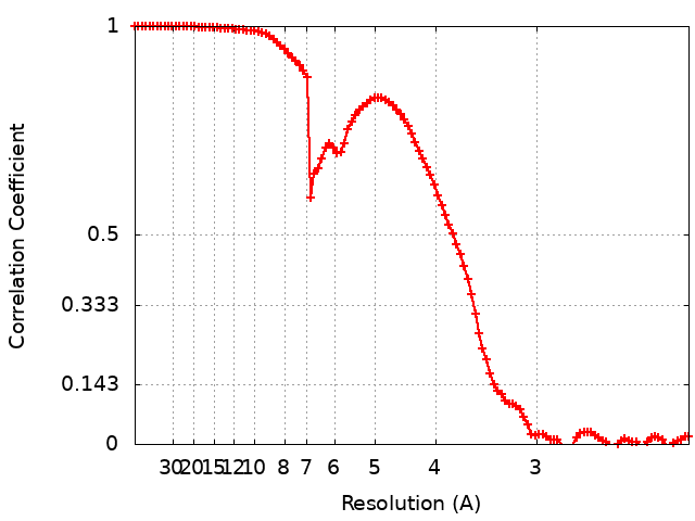 |
