 Movie
Movie Controller

[English] 日本語
 Yorodumi
- EMDB-2323: Electron cryo-microscopy of a cross-beta amyloid fibril polymorph -
+ Open data

- Basic information
| Entry | Database: EMDB / ID: EMD-2323 | |||||||||
|---|---|---|---|---|---|---|---|---|---|---|
| Title | Electron cryo-microscopy of a cross-beta amyloid fibril polymorph | |||||||||
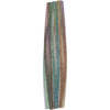
 Map data Map data | Reconstruction of triplet cross-beta amyloid fibril polymorph | |||||||||
 Sample Sample |
| |||||||||
 Keywords Keywords | amyloid fibrils / cross-beta structure / protein aggregation / polymorphism | |||||||||
| Function / homology |  Function and homology information Function and homology informationThe canonical retinoid cycle in rods (twilight vision) / Retinoid metabolism and transport / thyroid hormone metabolic process / hormone binding / Neutrophil degranulation / thyroid hormone binding / purine nucleobase metabolic process / hormone activity / protein-containing complex binding / protein-containing complex ...The canonical retinoid cycle in rods (twilight vision) / Retinoid metabolism and transport / thyroid hormone metabolic process / hormone binding / Neutrophil degranulation / thyroid hormone binding / purine nucleobase metabolic process / hormone activity / protein-containing complex binding / protein-containing complex / extracellular space / identical protein binding Similarity search - Function | |||||||||
| Biological species |  | |||||||||
| Method | single particle reconstruction / cryo EM / Resolution: 12.2 Å | |||||||||
 Authors Authors | Fitzpatrick AWP / Debelouchina GT / Bayro MJ / Clare DK / Caporini MA / Bajaj VS / Jaroniec CP / Wang L / Ladizhansky V / Muller S ...Fitzpatrick AWP / Debelouchina GT / Bayro MJ / Clare DK / Caporini MA / Bajaj VS / Jaroniec CP / Wang L / Ladizhansky V / Muller S / MacPhee CE / Waudby CA / Mott HR / De-Simone A / Knowles TPJ / Saibil HR / Vendruscolo M / Orlova EV / Griffin RG / Dobson CM | |||||||||
 Citation Citation | Journal: Proc Natl Acad Sci U S A / Year: 2013 Title: Atomic structure and hierarchical assembly of a cross-β amyloid fibril. Authors: Anthony W P Fitzpatrick / Galia T Debelouchina / Marvin J Bayro / Daniel K Clare / Marc A Caporini / Vikram S Bajaj / Christopher P Jaroniec / Luchun Wang / Vladimir Ladizhansky / Shirley A ...Authors: Anthony W P Fitzpatrick / Galia T Debelouchina / Marvin J Bayro / Daniel K Clare / Marc A Caporini / Vikram S Bajaj / Christopher P Jaroniec / Luchun Wang / Vladimir Ladizhansky / Shirley A Müller / Cait E MacPhee / Christopher A Waudby / Helen R Mott / Alfonso De Simone / Tuomas P J Knowles / Helen R Saibil / Michele Vendruscolo / Elena V Orlova / Robert G Griffin / Christopher M Dobson /  Abstract: The cross-β amyloid form of peptides and proteins represents an archetypal and widely accessible structure consisting of ordered arrays of β-sheet filaments. These complex aggregates have ...The cross-β amyloid form of peptides and proteins represents an archetypal and widely accessible structure consisting of ordered arrays of β-sheet filaments. These complex aggregates have remarkable chemical and physical properties, and the conversion of normally soluble functional forms of proteins into amyloid structures is linked to many debilitating human diseases, including several common forms of age-related dementia. Despite their importance, however, cross-β amyloid fibrils have proved to be recalcitrant to detailed structural analysis. By combining structural constraints from a series of experimental techniques spanning five orders of magnitude in length scale--including magic angle spinning nuclear magnetic resonance spectroscopy, X-ray fiber diffraction, cryoelectron microscopy, scanning transmission electron microscopy, and atomic force microscopy--we report the atomic-resolution (0.5 Å) structures of three amyloid polymorphs formed by an 11-residue peptide. These structures reveal the details of the packing interactions by which the constituent β-strands are assembled hierarchically into protofilaments, filaments, and mature fibrils. | |||||||||
| History |
|
- Structure visualization
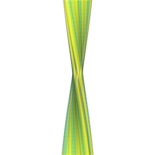
| Movie |
Movie viewer |
|---|---|
| Structure viewer | EM map: SurfViewMolmilJmol/JSmol |
| Supplemental images |
- Downloads & links
-EMDB archive
| Map data | emd_2323.map.gz | 9.8 MB | EMDB map data format | |
|---|---|---|---|---|
| Header (meta data) | emd-2323-v30.xmlemd-2323.xml | 10.6 KB 10.6 KB | Display Display | EMDB header |
| Images | 2323.tif | 732.8 KB | ||
| Archive directory |  http://ftp.pdbj.org/pub/emdb/structures/EMD-2323ftp://ftp.pdbj.org/pub/emdb/structures/EMD-2323 http://ftp.pdbj.org/pub/emdb/structures/EMD-2323ftp://ftp.pdbj.org/pub/emdb/structures/EMD-2323 | HTTPS FTP |
-Validation report
| Summary document | emd_2323_validation.pdf.gz | 259.3 KB | Display | EMDB validaton report |
|---|---|---|---|---|
| Full document | emd_2323_full_validation.pdf.gz | 258.9 KB | Display | |
| Data in XML | emd_2323_validation.xml.gz | 4.9 KB | Display | |
| Arichive directory | https://ftp.pdbj.org/pub/emdb/validation_reports/EMD-2323ftp://ftp.pdbj.org/pub/emdb/validation_reports/EMD-2323 | HTTPS FTP |
-Related structure data
| Related structure data | 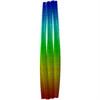 2m5mMC 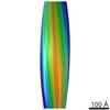 2324C 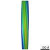 5590C 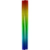 2m5kC 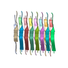 2m5nC 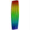 3zpkC M: atomic model generated by this map C: citing same article ( |
|---|---|
| Similar structure data |

-Links
| EMDB pages | EMDB (EBI/PDBe) / EMDataResource |
|---|---|
| Related items in Molecule of the Month |


-Map
| File | Download / File: emd_2323.map.gz / Format: CCP4 / Size: 16 MB / Type: IMAGE STORED AS FLOATING POINT NUMBER (4 BYTES) | ||||||||||||||||||||||||||||||||||||||||||||||||||||||||||||||||||||
|---|---|---|---|---|---|---|---|---|---|---|---|---|---|---|---|---|---|---|---|---|---|---|---|---|---|---|---|---|---|---|---|---|---|---|---|---|---|---|---|---|---|---|---|---|---|---|---|---|---|---|---|---|---|---|---|---|---|---|---|---|---|---|---|---|---|---|---|---|---|
| Annotation | Reconstruction of triplet cross-beta amyloid fibril polymorph | ||||||||||||||||||||||||||||||||||||||||||||||||||||||||||||||||||||
| Voxel size | X=Y=Z: 1.8 Å | ||||||||||||||||||||||||||||||||||||||||||||||||||||||||||||||||||||
| Density |
| ||||||||||||||||||||||||||||||||||||||||||||||||||||||||||||||||||||
| Symmetry | Space group: 1 | ||||||||||||||||||||||||||||||||||||||||||||||||||||||||||||||||||||
| Details | EMDB XML:
CCP4 map header:
| ||||||||||||||||||||||||||||||||||||||||||||||||||||||||||||||||||||
-Supplemental data
- Sample components
-Entire : Triplet cross-beta amyloid fibril polymorph
| Entire | Name: Triplet cross-beta amyloid fibril polymorph |
|---|---|
| Components |
|
-Supramolecule #1000: Triplet cross-beta amyloid fibril polymorph
| Supramolecule | Name: Triplet cross-beta amyloid fibril polymorph / type: sample / ID: 1000 / Number unique components: 1 |
|---|
-Macromolecule #1: Transthyretin(105-115)
| Macromolecule | Name: Transthyretin(105-115) / type: protein_or_peptide / ID: 1 / Name.synonym: TTR(105-115) / Recombinant expression: No |
|---|---|
| Source (natural) | Organism: |
| Sequence | UniProtKB: Transthyretin |
-Experimental details
-Structure determination
| Method | cryo EM |
|---|---|
 Processing Processing | single particle reconstruction |
| Aggregation state | filament |
-Sample preparation
| Concentration | 1 mg/mL |
|---|---|
| Buffer | pH: 2 / Details: 10% acetonitrile/water solution |
| Grid | Details: For cryo-EM imaging, fibrils were applied to holey carbon films (R2/2, QuantifoilMicro Tools GmbH, Jena, Germany) that were immediately plunge-frozen at liquid nitrogen temperature. |
| Vitrification | Cryogen name: ETHANE / Chamber humidity: 95 % / Chamber temperature: 99 K / Instrument: HOMEMADE PLUNGER Method: For cryo-EM imaging, fibrils were applied to holey carbon films (R2/2, QuantifoilMicro Tools GmbH, Jena, Germany) that were immediately plunge-frozen at liquid nitrogen temperature. |
- Electron microscopy
| Microscope | FEI TECNAI F20 |
|---|---|
| Temperature | Min: 99 K / Max: 100 K / Average: 99 K |
| Alignment procedure | Legacy - Astigmatism: Objective lens astigmatism was corrected at 100,000 times magnification. |
| Date | Mar 3, 2006 |
| Image recording | Category: FILM / Film or detector model: KODAK SO-163 FILM / Digitization - Scanner: ZEISS SCAI / Number real images: 250 |
| Electron beam | Acceleration voltage: 200 kV / Electron source:  FIELD EMISSION GUN FIELD EMISSION GUN |
| Electron optics | Calibrated magnification: 40000 / Illumination mode: FLOOD BEAM / Imaging mode: BRIGHT FIELD / Nominal defocus max: 3.0 µm / Nominal defocus min: 0.9 µm / Nominal magnification: 40000 |
| Sample stage | Specimen holder: Nitrogen cooled / Specimen holder model: GATAN LIQUID NITROGEN |
| Experimental equipment |  Model: Tecnai F20 / Image courtesy: FEI Company |
-Image processing
| Details | The particles were selected using an automatic selection program. |
|---|---|
| CTF correction | Details: CTFFIND3 |
| Final reconstruction | Applied symmetry - Point group: C1 (asymmetric) / Algorithm: OTHER / Resolution.type: BY AUTHOR / Resolution: 12.2 Å / Resolution method: FSC 0.5 CUT-OFF / Software - Name: SPIER, IMAGIC, BKRP / Number images used: 152 |
| Final two d classification | Number classes: 28 |
