 Movie
Movie Controller


+ Open data

- Basic information
| Entry | Database: EMDB / ID: EMD-23059 | |||||||||
|---|---|---|---|---|---|---|---|---|---|---|
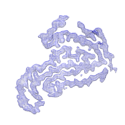
| Title | TDP-43 LCD amyloid fibrils | |||||||||
 Map data Map data | ||||||||||
 Sample Sample |
| |||||||||
 Keywords Keywords |  TDP-43 / amyloid / neurodegenerative diseases / amyotropic lateral sclerosis / PROTEIN FIBRIL TDP-43 / amyloid / neurodegenerative diseases / amyotropic lateral sclerosis / PROTEIN FIBRIL | |||||||||
| Function / homology |  Function and homology information Function and homology informationnuclear inner membrane organization / interchromatin granule / perichromatin fibrils / 3'-UTR-mediated mRNA destabilization / 3'-UTR-mediated mRNA stabilization / intracellular non-membrane-bounded organelle / negative regulation by host of viral transcription / pre-mRNA intronic binding / response to endoplasmic reticulum stress / RNA splicing ...nuclear inner membrane organization / interchromatin granule / perichromatin fibrils / 3'-UTR-mediated mRNA destabilization / 3'-UTR-mediated mRNA stabilization / intracellular non-membrane-bounded organelle / negative regulation by host of viral transcription / pre-mRNA intronic binding / response to endoplasmic reticulum stress / RNA splicing / negative regulation of protein phosphorylation / molecular condensate scaffold activity / mRNA 3'-UTR binding / regulation of protein stability / regulation of circadian rhythm / positive regulation of insulin secretion / mRNA processing / cytoplasmic stress granule / positive regulation of protein import into nucleus / rhythmic process / regulation of gene expression / double-stranded DNA binding / regulation of apoptotic process / amyloid fibril formation / regulation of cell cycle / nuclear speck / RNA polymerase II cis-regulatory region sequence-specific DNA binding / negative regulation of gene expression / lipid binding / mitochondrion / DNA binding / RNA binding / nucleoplasm / identical protein binding / nucleusSimilarity search - Function | |||||||||
| Biological species |  Homo sapiens (human) Homo sapiens (human) | |||||||||
| Method | helical reconstruction / cryo EM / Resolution: 3.2 Å | |||||||||
 Authors Authors | Li Q / Babinchak WM | |||||||||
| Funding support |  United States, 2 items United States, 2 items
| |||||||||
 Citation Citation | Journal: Nat Commun / Year: 2021 Title: Cryo-EM structure of amyloid fibrils formed by the entire low complexity domain of TDP-43. Authors: Qiuye Li / W Michael Babinchak / Witold K Surewicz / Abstract: Amyotrophic lateral sclerosis and several other neurodegenerative diseases are associated with brain deposits of amyloid-like aggregates formed by the C-terminal fragments of TDP-43 that contain the ...Amyotrophic lateral sclerosis and several other neurodegenerative diseases are associated with brain deposits of amyloid-like aggregates formed by the C-terminal fragments of TDP-43 that contain the low complexity domain of the protein. Here, we report the cryo-EM structure of amyloid formed from the entire TDP-43 low complexity domain in vitro at pH 4. This structure reveals single protofilament fibrils containing a large (139-residue), tightly packed core. While the C-terminal part of this core region is largely planar and characterized by a small proportion of hydrophobic amino acids, the N-terminal region contains numerous hydrophobic residues and has a non-planar backbone conformation, resulting in rugged surfaces of fibril ends. The structural features found in these fibrils differ from those previously found for fibrils generated from short protein fragments. The present atomic model for TDP-43 LCD fibrils provides insight into potential structural perturbations caused by phosphorylation and disease-related mutations. | |||||||||
| History |
|
- Structure visualization
| Movie |
Movie viewer |
|---|---|
| Structure viewer | EM map: SurfViewMolmilJmol/JSmol |
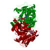
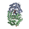
| Supplemental images |
- Downloads & links
-EMDB archive
| Map data | emd_23059.map.gz | 2 MB | EMDB map data format | |
|---|---|---|---|---|
| Header (meta data) | emd-23059-v30.xmlemd-23059.xml | 9 KB 9 KB | Display Display | EMDB header |
| Images |  emd_23059.png emd_23059.png | 125.5 KB | ||
| Filedesc metadata | emd-23059.cif.gz | 4.8 KB | ||
| Archive directory |  http://ftp.pdbj.org/pub/emdb/structures/EMD-23059ftp://ftp.pdbj.org/pub/emdb/structures/EMD-23059 http://ftp.pdbj.org/pub/emdb/structures/EMD-23059ftp://ftp.pdbj.org/pub/emdb/structures/EMD-23059 | HTTPS FTP |
-Related structure data
| Related structure data | 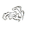 7kwzMC M: atomic model generated by this map C: citing same article ( |
|---|---|
| Similar structure data |
-Links
| EMDB pages | EMDB (EBI/PDBe) / EMDataResource |
|---|---|
| Related items in Molecule of the Month |

-Map
| File | Download / File: emd_23059.map.gz / Format: CCP4 / Size: 30.5 MB / Type: IMAGE STORED AS FLOATING POINT NUMBER (4 BYTES) | ||||||||||||||||||||||||||||||||||||||||||||||||||||||||||||
|---|---|---|---|---|---|---|---|---|---|---|---|---|---|---|---|---|---|---|---|---|---|---|---|---|---|---|---|---|---|---|---|---|---|---|---|---|---|---|---|---|---|---|---|---|---|---|---|---|---|---|---|---|---|---|---|---|---|---|---|---|---|
| Voxel size | X=Y=Z: 0.828 Å | ||||||||||||||||||||||||||||||||||||||||||||||||||||||||||||
| Density |
| ||||||||||||||||||||||||||||||||||||||||||||||||||||||||||||
| Symmetry | Space group: 1 | ||||||||||||||||||||||||||||||||||||||||||||||||||||||||||||
| Details | EMDB XML:
CCP4 map header:
| ||||||||||||||||||||||||||||||||||||||||||||||||||||||||||||
-Supplemental data
- Sample components
-Entire : Amyloid fibrils formed by TDP-43 low complexity domain
| Entire | Name: Amyloid fibrils formed by TDP-43 low complexity domain |
|---|---|
| Components |
|
-Supramolecule #1: Amyloid fibrils formed by TDP-43 low complexity domain
| Supramolecule | Name: Amyloid fibrils formed by TDP-43 low complexity domain type: complex / ID: 1 / Parent: 0 / Macromolecule list: all |
|---|---|
| Source (natural) | Organism: Homo sapiens (human) |
-Macromolecule #1: Isoform 2 of TAR DNA-binding protein 43
| Macromolecule | Name: Isoform 2 of TAR DNA-binding protein 43 / type: protein_or_peptide / ID: 1 / Number of copies: 5 / Enantiomer: LEVO |
|---|---|
| Source (natural) | Organism: Homo sapiens (human) |
| Molecular weight | Theoretical: 14.570482 KDa |
| Recombinant expression | Organism:  Escherichia coli (E. coli) Escherichia coli (E. coli) |
| Sequence | String: NRQLERSGRF GGNPGGFGNQ GGFGNSRGGG AGLGNNQGSN MGGGMNFGAF SINPAMMAAA QAALQSSWGM MGMLASQQNQ SGPSGNNQN QGNMQREPNQ AFGSGNNSYS GSNSGAAIGW GSASNAGSGS GFNGGFGSSM DSKSSGWGM UniProtKB: TAR DNA-binding protein 43 |
-Experimental details
-Structure determination
| Method | cryo EM |
|---|---|
 Processing Processing | helical reconstruction |
| Aggregation state | filament |
-Sample preparation
| Buffer | pH: 4 |
|---|---|
| Vitrification | Cryogen name: ETHANE |
- Electron microscopy
| Microscope | FEI TITAN KRIOS |
|---|---|
| Electron beam | Acceleration voltage: 300 kV / Electron source: FIELD EMISSION GUN |
| Electron optics | Illumination mode: OTHER / Imaging mode: OTHER |
| Image recording | Film or detector model: GATAN K3 BIOQUANTUM (6k x 4k) / Average electron dose: 42.0 e/Å2 |
| Experimental equipment |  Model: Titan Krios / Image courtesy: FEI Company |
-Image processing
| Startup model | Type of model: NONE |
|---|---|
| Final angle assignment | Type: NOT APPLICABLE |
| Final reconstruction | Applied symmetry - Helical parameters - Δz: 4.73 Å Applied symmetry - Helical parameters - Δ&Phi: -1.66 ° Applied symmetry - Helical parameters - Axial symmetry: C1 (asymmetric) Resolution.type: BY AUTHOR / Resolution: 3.2 Å / Resolution method: FSC 0.143 CUT-OFF / Number images used: 11026 |
