National Institutes of Health/Eunice Kennedy Shriver National Institute of Child Health & Human Development (NIH/NICHD)
R01 GM61576
United States
Citation
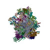
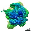
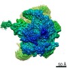
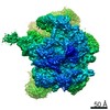
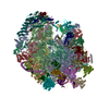
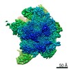
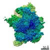
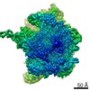
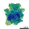
Journal: Nat Commun / Year: 2020 Title: Structures of the human mitochondrial ribosome bound to EF-G1 reveal distinct features of mitochondrial translation elongation. Authors: Ravi Kiran Koripella / Manjuli R Sharma / Kalpana Bhargava / Partha P Datta / Prem S Kaushal / Pooja Keshavan / Linda L Spremulli / Nilesh K Banavali / Rajendra K Agrawal / Abstract: The mammalian mitochondrial ribosome (mitoribosome) and its associated translational factors have evolved to accommodate greater participation of proteins in mitochondrial translation. Here we ...The mammalian mitochondrial ribosome (mitoribosome) and its associated translational factors have evolved to accommodate greater participation of proteins in mitochondrial translation. Here we present the 2.68-3.96 Å cryo-EM structures of the human 55S mitoribosome in complex with the human mitochondrial elongation factor G1 (EF-G1) in three distinct conformational states, including an intermediate state and a post-translocational state. These structures reveal the role of several mitochondria-specific (mito-specific) mitoribosomal proteins (MRPs) and a mito-specific segment of EF-G1 in mitochondrial tRNA (tRNA) translocation. In particular, the mito-specific C-terminal extension in EF-G1 is directly involved in translocation of the acceptor arm of the A-site tRNA. In addition to the ratchet-like and independent head-swiveling motions exhibited by the small mitoribosomal subunit, we discover significant conformational changes in MRP mL45 at the nascent polypeptide-exit site within the large mitoribosomal subunit that could be critical for tethering of the elongating mitoribosome onto the inner-mitochondrial membrane.
History
Deposition
Jun 23, 2020
-
Header (metadata) release
Aug 5, 2020
-
Map release
Aug 5, 2020
-
Update
Aug 12, 2020
-
Current status
Aug 12, 2020
Processing site: RCSB / Status: Released
-
Structure visualization
Movie
Surface view with section colored by density value
Acceleration voltage: 300 kV / Electron source: OTHER
Electron optics
Illumination mode: OTHER / Imaging mode: OTHER
Image recording
#0 - Image recording ID: 1 / #0 - Film or detector model: GATAN K2 SUMMIT (4k x 4k) / #0 - Average electron dose: 69.21 e/Å2 / #1 - Image recording ID: 2 #1 - Film or detector model: DIRECT ELECTRON DE-16 (4k x 4k) #1 - Detector mode: COUNTING / #1 - Average electron dose: 50.0 e/Å2
Experimental equipment
Model: Tecnai Polara / Image courtesy: FEI Company
In the structure databanks used in Yorodumi, some data are registered as the other names, "COVID-19 virus" and "2019-nCoV". Here are the details of the virus and the list of structure data.
Jan 31, 2019. EMDB accession codes are about to change! (news from PDBe EMDB page)
EMDB accession codes are about to change! (news from PDBe EMDB page)
The allocation of 4 digits for EMDB accession codes will soon come to an end. Whilst these codes will remain in use, new EMDB accession codes will include an additional digit and will expand incrementally as the available range of codes is exhausted. The current 4-digit format prefixed with “EMD-” (i.e. EMD-XXXX) will advance to a 5-digit format (i.e. EMD-XXXXX), and so on. It is currently estimated that the 4-digit codes will be depleted around Spring 2019, at which point the 5-digit format will come into force.
The EM Navigator/Yorodumi systems omit the EMD- prefix.
Related info.:Q: What is EMD? / ID/Accession-code notation in Yorodumi/EM Navigator
Yorodumi is a browser for structure data from EMDB, PDB, SASBDB, etc.
This page is also the successor to EM Navigator detail page, and also detail information page/front-end page for Omokage search.
The word "yorodu" (or yorozu) is an old Japanese word meaning "ten thousand". "mi" (miru) is to see.
Related info.:EMDB / PDB / SASBDB / Comparison of 3 databanks / Yorodumi Search / Aug 31, 2016. New EM Navigator & Yorodumi / Yorodumi Papers / Jmol/JSmol / Function and homology information / Changes in new EM Navigator and Yorodumi
 Movie
Movie Controller


 Open data
 Basic information
 Map data
 Sample
 Bos taurus (cattle)
 Authors
 United States, 1 items
  Citation
 Structure visualization
 Movie viewer
 Downloads & links
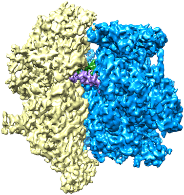 emd_22209.png
 http://ftp.pdbj.org/pub/emdb/structures/EMD-22209


 Sample components
 Processing
 Electron microscopy