 Movie
Movie Controller

[English] 日本語
 Yorodumi
- EMDB-21983: Single-Particle Cryo-EM Structure of Arabinosyltransferase EmbB f... -
+ Open data

- Basic information
| Entry | Database: EMDB / ID: EMD-21983 | |||||||||||||||||||||||||||||||||||||||
|---|---|---|---|---|---|---|---|---|---|---|---|---|---|---|---|---|---|---|---|---|---|---|---|---|---|---|---|---|---|---|---|---|---|---|---|---|---|---|---|---|
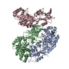
| Title | Single-Particle Cryo-EM Structure of Arabinosyltransferase EmbB from Mycobacterium smegmatis | |||||||||||||||||||||||||||||||||||||||
 Map data Map data | Density modified map | |||||||||||||||||||||||||||||||||||||||
 Sample Sample |
| |||||||||||||||||||||||||||||||||||||||
 Keywords Keywords |  Glycosyltransferase / membrane protein / nanodisc Glycosyltransferase / membrane protein / nanodisc | |||||||||||||||||||||||||||||||||||||||
| Function / homology |  Function and homology informationindolylacetylinositol arabinosyltransferase / indolylacetylinositol arabinosyltransferase activity / arabinosyltransferase activity / Actinobacterium-type cell wall biogenesis / Transferases; Glycosyltransferases; Pentosyltransferases / cell wall organization / plasma membrane Function and homology informationindolylacetylinositol arabinosyltransferase / indolylacetylinositol arabinosyltransferase activity / arabinosyltransferase activity / Actinobacterium-type cell wall biogenesis / Transferases; Glycosyltransferases; Pentosyltransferases / cell wall organization / plasma membraneSimilarity search - Function | |||||||||||||||||||||||||||||||||||||||
| Biological species |  Mycolicibacterium smegmatis (bacteria) / Mycolicibacterium smegmatis (strain ATCC 700084 / mc(2)155) (bacteria) Mycolicibacterium smegmatis (bacteria) / Mycolicibacterium smegmatis (strain ATCC 700084 / mc(2)155) (bacteria) | |||||||||||||||||||||||||||||||||||||||
| Method | single particle reconstruction / cryo EM / Resolution: 3.3 Å | |||||||||||||||||||||||||||||||||||||||
 Authors Authors | Tan YZ / Rodrigues J | |||||||||||||||||||||||||||||||||||||||
| Funding support |  United States, United States,  Portugal, European Union, Portugal, European Union,  Canada, 12 items Canada, 12 items
| |||||||||||||||||||||||||||||||||||||||
 Citation Citation | Journal: Nat Commun / Year: 2020 Title: Cryo-EM structure of arabinosyltransferase EmbB from Mycobacterium smegmatis. Authors: Yong Zi Tan / José Rodrigues / James E Keener / Ruixiang Blake Zheng / Richard Brunton / Brian Kloss / Sabrina I Giacometti / Ana L Rosário / Lei Zhang / Michael Niederweis / Oliver B ...Authors: Yong Zi Tan / José Rodrigues / James E Keener / Ruixiang Blake Zheng / Richard Brunton / Brian Kloss / Sabrina I Giacometti / Ana L Rosário / Lei Zhang / Michael Niederweis / Oliver B Clarke / Todd L Lowary / Michael T Marty / Margarida Archer / Clinton S Potter / Bridget Carragher / Filippo Mancia /  Abstract: Arabinosyltransferase B (EmbB) belongs to a family of membrane-bound glycosyltransferases that build the lipidated polysaccharides of the mycobacterial cell envelope, and are targets of anti- ...Arabinosyltransferase B (EmbB) belongs to a family of membrane-bound glycosyltransferases that build the lipidated polysaccharides of the mycobacterial cell envelope, and are targets of anti-tuberculosis drug ethambutol. We present the 3.3 Å resolution single-particle cryo-electron microscopy structure of Mycobacterium smegmatis EmbB, providing insights on substrate binding and reaction mechanism. Mutations that confer ethambutol resistance map mostly around the putative active site, suggesting this to be the location of drug binding. | |||||||||||||||||||||||||||||||||||||||
| History |
|
- Structure visualization
| Movie |
Movie viewer |
|---|---|
| Structure viewer | EM map: SurfViewMolmilJmol/JSmol |
| Supplemental images |

- Downloads & links
-EMDB archive
| Map data | emd_21983.map.gz | 59.7 MB | EMDB map data format | |
|---|---|---|---|---|
| Header (meta data) | emd-21983-v30.xmlemd-21983.xml | 37.2 KB 37.2 KB | Display Display | EMDB header |
| FSC (resolution estimation) | emd_21983_fsc.xml | 9.8 KB | Display | FSC data file |
| Images | 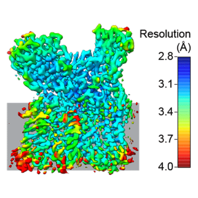 emd_21983.png emd_21983.png | 166.3 KB | ||
| Masks | emd_21983_msk_1.map | 64 MB | Mask map | |
| Filedesc metadata | emd-21983.cif.gz | 7.6 KB | ||
| Others | emd_21983_additional_1.map.gzemd_21983_additional_2.map.gzemd_21983_additional_3.map.gzemd_21983_additional_4.map.gzemd_21983_additional_5.map.gzemd_21983_additional_6.map.gzemd_21983_half_map_1.map.gzemd_21983_half_map_2.map.gz | 18.2 MB 59.6 MB 8.4 MB 7.1 MB 107.1 KB 3.4 MB 59.4 MB 59.4 MB | ||
| Archive directory |  http://ftp.pdbj.org/pub/emdb/structures/EMD-21983ftp://ftp.pdbj.org/pub/emdb/structures/EMD-21983 http://ftp.pdbj.org/pub/emdb/structures/EMD-21983ftp://ftp.pdbj.org/pub/emdb/structures/EMD-21983 | HTTPS FTP |
-Related structure data
| Related structure data |  6x0oMC M: atomic model generated by this map C: citing same article ( |
|---|---|
| Similar structure data | |
| EM raw data | EMPIAR-10420 (Title: Single-Particle Cryo-EM of Arabinosyltransferase EmbB from Mycobacterium smegmatis, Collected using both Falcon III and K2 Detectors Data size: 4.5 TB Data #1: Unaligned and compressed multiframe movies from Falcon III Detector [micrographs - multiframe] Data #2: Unaligned and compressed multiframe movies from K2 Summit Detector [micrographs - multiframe] Data #3: Aligned and dose-weighted micrographs from Falcon III Detector [micrographs - single frame] Data #4: Aligned and dose-weighted micrographs from K2 Summit Detector [micrographs - single frame] Data #5: Final Particle Stack with Refined Euler Angles and Shifts [picked particles - single frame - processed]) |


-Links
| EMDB pages | EMDB (EBI/PDBe) / EMDataResource |
|---|
-Map
| File | Download / File: emd_21983.map.gz / Format: CCP4 / Size: 64 MB / Type: IMAGE STORED AS FLOATING POINT NUMBER (4 BYTES) | ||||||||||||||||||||||||||||||||||||||||||||||||||||||||||||||||||||
|---|---|---|---|---|---|---|---|---|---|---|---|---|---|---|---|---|---|---|---|---|---|---|---|---|---|---|---|---|---|---|---|---|---|---|---|---|---|---|---|---|---|---|---|---|---|---|---|---|---|---|---|---|---|---|---|---|---|---|---|---|---|---|---|---|---|---|---|---|---|
| Annotation | Density modified map | ||||||||||||||||||||||||||||||||||||||||||||||||||||||||||||||||||||
| Voxel size | X=Y=Z: 1 Å | ||||||||||||||||||||||||||||||||||||||||||||||||||||||||||||||||||||
| Density |
| ||||||||||||||||||||||||||||||||||||||||||||||||||||||||||||||||||||
| Symmetry | Space group: 1 | ||||||||||||||||||||||||||||||||||||||||||||||||||||||||||||||||||||
| Details | EMDB XML:
CCP4 map header:
| ||||||||||||||||||||||||||||||||||||||||||||||||||||||||||||||||||||
-Supplemental data
-Mask #1
| File | emd_21983_msk_1.map | ||||||||||||
|---|---|---|---|---|---|---|---|---|---|---|---|---|---|
| Projections & Slices |
| ||||||||||||
| Density Histograms |
 Z
Z Y
Y X
X


-Additional map: Raw map
| File | emd_21983_additional_1.map | ||||||||||||
|---|---|---|---|---|---|---|---|---|---|---|---|---|---|
| Annotation | Raw map | ||||||||||||
| Projections & Slices |
| ||||||||||||
| Density Histograms |


-Additional map: CryoSPARC Sharpened Map
| File | emd_21983_additional_2.map | ||||||||||||
|---|---|---|---|---|---|---|---|---|---|---|---|---|---|
| Annotation | CryoSPARC Sharpened Map | ||||||||||||
| Projections & Slices |
| ||||||||||||
| Density Histograms |


-Additional map: 3DFSC - Raw
| File | emd_21983_additional_3.map | ||||||||||||
|---|---|---|---|---|---|---|---|---|---|---|---|---|---|
| Annotation | 3DFSC - Raw | ||||||||||||
| Projections & Slices |
| ||||||||||||
| Density Histograms |


-Additional map: 3DFSC - Thresholded
| File | emd_21983_additional_4.map | ||||||||||||
|---|---|---|---|---|---|---|---|---|---|---|---|---|---|
| Annotation | 3DFSC - Thresholded | ||||||||||||
| Projections & Slices |
| ||||||||||||
| Density Histograms |


-Additional map: 3DFSC - Thresholded and binarized
| File | emd_21983_additional_5.map | ||||||||||||
|---|---|---|---|---|---|---|---|---|---|---|---|---|---|
| Annotation | 3DFSC - Thresholded and binarized | ||||||||||||
| Projections & Slices |
| ||||||||||||
| Density Histograms |


-Additional map: Local resolution map
| File | emd_21983_additional_6.map | ||||||||||||
|---|---|---|---|---|---|---|---|---|---|---|---|---|---|
| Annotation | Local resolution map | ||||||||||||
| Projections & Slices |
| ||||||||||||
| Density Histograms |


-Half map: Half map 1
| File | emd_21983_half_map_1.map | ||||||||||||
|---|---|---|---|---|---|---|---|---|---|---|---|---|---|
| Annotation | Half map 1 | ||||||||||||
| Projections & Slices |
| ||||||||||||
| Density Histograms |


-Half map: Half map 2
| File | emd_21983_half_map_2.map | ||||||||||||
|---|---|---|---|---|---|---|---|---|---|---|---|---|---|
| Annotation | Half map 2 | ||||||||||||
| Projections & Slices |
| ||||||||||||
| Density Histograms |


- Sample components
-Entire : Integral membrane indolylacetylinositol arabinosyltransferase EmbB
| Entire | Name: Integral membrane indolylacetylinositol arabinosyltransferase EmbB |
|---|---|
| Components |
|
-Supramolecule #1: Integral membrane indolylacetylinositol arabinosyltransferase EmbB
| Supramolecule | Name: Integral membrane indolylacetylinositol arabinosyltransferase EmbB type: organelle_or_cellular_component / ID: 1 / Parent: 0 / Macromolecule list: #1 |
|---|---|
| Source (natural) | Organism: Mycolicibacterium smegmatis (bacteria) |
| Molecular weight | Theoretical: 119 KDa |
-Macromolecule #1: Integral membrane indolylacetylinositol arabinosyltransferase EmbB
| Macromolecule | Name: Integral membrane indolylacetylinositol arabinosyltransferase EmbB type: protein_or_peptide / ID: 1 / Number of copies: 1 / Enantiomer: LEVO / EC number: indolylacetylinositol arabinosyltransferase |
|---|---|
| Source (natural) | Organism: Mycolicibacterium smegmatis (strain ATCC 700084 / mc(2)155) (bacteria) Strain: ATCC 700084 / mc(2)155 |
| Molecular weight | Theoretical: 118.835375 KDa |
| Recombinant expression | Organism: Escherichia coli (E. coli) |
| Sequence | String: MSGNMDEAVS GNMDEAVSAG KDVRIARWVA TIAGLLGFVL SVSIPLLPVT QTTATLNWPQ QGRLDNVTAP LISQAPLELT ATVPCSVVR DLPPEGGLVF GTAPAEGRDA ALNAMLVNVT ETRVDVIVRN VVVASVNRDR VAGPDCQRIE ITSNLDGTYA D FVGLTQIS ...String: MSGNMDEAVS GNMDEAVSAG KDVRIARWVA TIAGLLGFVL SVSIPLLPVT QTTATLNWPQ QGRLDNVTAP LISQAPLELT ATVPCSVVR DLPPEGGLVF GTAPAEGRDA ALNAMLVNVT ETRVDVIVRN VVVASVNRDR VAGPDCQRIE ITSNLDGTYA D FVGLTQIS GEDAGKLQRT GYPDPNLRPA IVGVFTDLTG PAPQGLSVSA EIDTRFTTHP TALKLAAMLL AIVSTVIALL AL WRLDRLD GRRMHRLIPT RWRTVTAVDG VVVGGMAIWY VIGANSSDDG YILQMARTAE HAGYMANYFR WFGSPEDPFG WYY NVLALM TKVSDASIWI RLPDLICALI CWLLLSREVL PRLGPAVAGS RAAMWAAGLV LLGAWMPFNN GLRPEGQIAT GALI TYVLI ERAVTSGRLT PAALAITTAA FTLGIQPTGL IAVAALLAGG RPILRIVMRR RRLVGTWPLI APLLAAGTVI LAVVF ADQT IATVLEATRI RTAIGPSQEW WTENLRYYYL ILPTTDGAIS RRVAFVFTAM CLFPSLFMML RRKHIAGVAR GPAWRL MGI IFATMFFLMF TPTKWIHHFG LFAAVGGAMA ALATVLVSPT VLRSARNRMA FLSLVLFVLA FCFASTNGWW YVSNFGA PF NNSVPKVGGV QISAIFFALS AIAALWAFWL HLTRRTESRV VDRLTAAPIP VAAGFMVVVM MASMAIGVVR QYPTYSNG W ANIRAFAGGC GLADDVLVEP DSNAGFLTPL PGAYGPLGPL GGEDPQGFSP DGVPDRIIAE AIRLNNPQPG TDYDWNRPI KLDEPGINGS TVPLPYGLDP KRVPVAGTYS TEAQQESRLS SAWYELPARD ETERAAHPLV VITAAGTITG ESVANGLTTG QTVDLEYAT RGPDGTLVPA GRVTPYDVGP TPSWRNLRYP RSEIPDDAVA VRVVAEDLSL SQGDWIAVTP PRVPELQSVQ E YVGSDQPV LMDWAVGLAF PCQQPMLHAN GVTEVPKFRI SPDYYAKLQS TDTWQDGING GLLGITDLLL RASVMSTYLS QD WGQDWGS LRKFDTVVEA TPAELDFGSQ THSGLYSPGP LRIRPAENLY FQGWSHPQFE K UniProtKB: Integral membrane indolylacetylinositol arabinosyltransferase EmbB |
-Macromolecule #2: CALCIUM ION
| Macromolecule | Name: CALCIUM ION / type: ligand / ID: 2 / Number of copies: 2 / Formula: CA |
|---|---|
| Molecular weight | Theoretical: 40.078 Da |
-Macromolecule #3: 1,2-DIPALMITOYL-PHOSPHATIDYL-GLYCEROLE
| Macromolecule | Name: 1,2-DIPALMITOYL-PHOSPHATIDYL-GLYCEROLE / type: ligand / ID: 3 / Number of copies: 2 / Formula: LHG |
|---|---|
| Molecular weight | Theoretical: 722.97 Da |
| Chemical component information |  ChemComp-LHG: |
-Experimental details
-Structure determination
| Method | cryo EM |
|---|---|
 Processing Processing | single particle reconstruction |
| Aggregation state | particle |
-Sample preparation
| Concentration | 8 mg/mL |
|---|---|
| Buffer | pH: 7.5 |
| Grid | Model: UltrAuFoil / Material: GOLD / Mesh: 300 / Support film - Material: GOLD / Support film - topology: HOLEY ARRAY / Pretreatment - Type: PLASMA CLEANING / Pretreatment - Time: 7 sec. / Pretreatment - Atmosphere: OTHER |
| Vitrification | Cryogen name: ETHANE / Chamber humidity: 80 % / Chamber temperature: 278 K / Instrument: LEICA EM GP |
- Electron microscopy
| Microscope | FEI TITAN KRIOS |
|---|---|
| Electron beam | Acceleration voltage: 300 kV / Electron source: FIELD EMISSION GUN |
| Electron optics | Illumination mode: FLOOD BEAM / Imaging mode: BRIGHT FIELDBright-field microscopy / Cs: 2.7 mm |
| Sample stage | Specimen holder model: FEI TITAN KRIOS AUTOGRID HOLDER / Cooling holder cryogen: NITROGEN |
| Image recording | #0 - Image recording ID: 1 / #0 - Film or detector model: FEI FALCON III (4k x 4k) / #0 - Detector mode: COUNTING / #0 - Number grids imaged: 1 / #0 - Number real images: 2158 / #0 - Average exposure time: 86.4 sec. / #0 - Average electron dose: 78.02 e/Å2 / #1 - Image recording ID: 2 / #1 - Film or detector model: GATAN K2 SUMMIT (4k x 4k) / #1 - Detector mode: COUNTING / #1 - Digitization - Dimensions - Width: 3838 pixel / #1 - Digitization - Dimensions - Height: 3710 pixel / #1 - Number grids imaged: 1 / #1 - Number real images: 7833 / #1 - Average exposure time: 8.0 sec. / #1 - Average electron dose: 77.53 e/Å2 |
| Experimental equipment |  Model: Titan Krios / Image courtesy: FEI Company |
-Image processing #1
| Particle selection | Number selected: 162271 |
|---|---|
| Startup model | Type of model: INSILICO MODEL / In silico model: Ab initio model / Details: Ab initio model was generated in CryoSPARC |
| Initial angle assignment | Type: MAXIMUM LIKELIHOOD / Software - Name: cryoSPARC (ver. 2) / Details: Non-uniform refinement in CryoSPARC was used. |
| Final angle assignment | Type: MAXIMUM LIKELIHOOD / Software - Name: cryoSPARC (ver. 2) / Details: Non-uniform refinement in CryoSPARC was used. |
| Final reconstruction | Applied symmetry - Point group: C1 (asymmetric) / Resolution.type: BY AUTHOR / Resolution: 3.3 Å / Resolution method: FSC 0.143 CUT-OFF / Software - Name: cryoSPARC (ver. 2) / Details: Combined with K2 dataset / Number images used: 57970 |
| Image processing ID | 1 |
| Image recording ID | 1 |
| FSC plot (resolution estimation) |  |
-Image processing #2
| Particle selection | Number selected: 700201 |
|---|---|
| Startup model | Type of model: INSILICO MODEL / In silico model: Ab initio model / Details: Ab initio model was generated in CryoSPARC |
| Initial angle assignment | Type: MAXIMUM LIKELIHOOD / Software - Name: cryoSPARC (ver. 2) / Details: Non-uniform refinement in CryoSPARC was used. |
| Final angle assignment | Type: MAXIMUM LIKELIHOOD / Software - Name: cryoSPARC (ver. 2) / Details: Non-uniform refinement in CryoSPARC was used. |
| Final reconstruction | Applied symmetry - Point group: C1 (asymmetric) / Resolution.type: BY AUTHOR / Resolution: 3.3 Å / Resolution method: FSC 0.143 CUT-OFF / Software - Name: cryoSPARC (ver. 2) / Details: Combined with Falcon III dataset / Number images used: 57970 |
| Image processing ID | 2 |
| Image recording ID | 2 |
| FSC plot (resolution estimation) | |

