 Movie
Movie Controller

[English] 日本語
 Yorodumi
- EMDB-21354: CryoEM Structure of the Plasmodium falciparum transporter PfFNT -
+ Open data

- Basic information
| Entry | Database: EMDB / ID: EMD-21354 | |||||||||
|---|---|---|---|---|---|---|---|---|---|---|
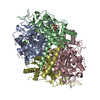
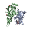
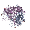
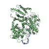
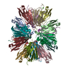
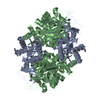
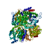
| Title | CryoEM Structure of the Plasmodium falciparum transporter PfFNT | |||||||||
 Map data Map data | ||||||||||
 Sample Sample |
| |||||||||
 Keywords Keywords |  Transporter / MEMBRANE PROTEIN Transporter / MEMBRANE PROTEIN | |||||||||
| Function / homology |  Function and homology information Function and homology informationhigh-affinity secondary active nitrite transmembrane transporter activity / lactate transmembrane transport / nitrite transport / lactate:proton symporter activity / membraneSimilarity search - Function | |||||||||
| Biological species |  Plasmodium falciparum 3D7 (eukaryote) / Plasmodium falciparum (isolate 3D7) (eukaryote) Plasmodium falciparum 3D7 (eukaryote) / Plasmodium falciparum (isolate 3D7) (eukaryote) | |||||||||
| Method | single particle reconstruction / cryo EM / Resolution: 2.56 Å | |||||||||
 Authors Authors | Su CC / Lyu M | |||||||||
 Citation Citation | Journal: EMBO Rep / Year: 2021 Title: Structural basis of transport and inhibition of the Plasmodium falciparum transporter PfFNT. Authors: Meinan Lyu / Chih-Chia Su / James W Kazura / Edward W Yu /  Abstract: The intra-erythrocyte stage of P. falciparum relies primarily on glycolysis to generate adenosine triphosphate (ATP) and the energy required to support growth and reproduction. Lactic acid, a ...The intra-erythrocyte stage of P. falciparum relies primarily on glycolysis to generate adenosine triphosphate (ATP) and the energy required to support growth and reproduction. Lactic acid, a metabolic byproduct of glycolysis, is potentially toxic as it lowers the pH inside the parasite. Plasmodium falciparum formate-nitrite transporter (PfFNT), a 34-kDa transmembrane protein, has been identified as a novel drug target as it exports lactate from inside the parasite to the surrounding parasitophorous vacuole within the erythrocyte cytosol. The structure and detailed molecular mechanism of this membrane protein are not yet available. Here we present structures of PfFNT in the absence and presence of the functional inhibitor MMV007839 at resolutions of 2.56 Å and 2.78 Å using single-particle cryo-electron microscopy. Genetic analysis and transport assay indicate that PfFNT is able to transfer lactate across the membrane. Combined, our data suggest a stepwise displacement mechanism for substrate transport. The PfFNT membrane protein is capable of picking up lactate ions from the parasite's cytosol, converting them to lactic acids and then exporting these acids into the extracellular space. | |||||||||
| History |
|
- Structure visualization
| Movie |
Movie viewer |
|---|---|
| Structure viewer | EM map: SurfViewMolmilJmol/JSmol |
| Supplemental images |

- Downloads & links
-EMDB archive
| Map data | emd_21354.map.gz | 2.5 MB | EMDB map data format | |
|---|---|---|---|---|
| Header (meta data) | emd-21354-v30.xmlemd-21354.xml | 9 KB 9 KB | Display Display | EMDB header |
| Images | 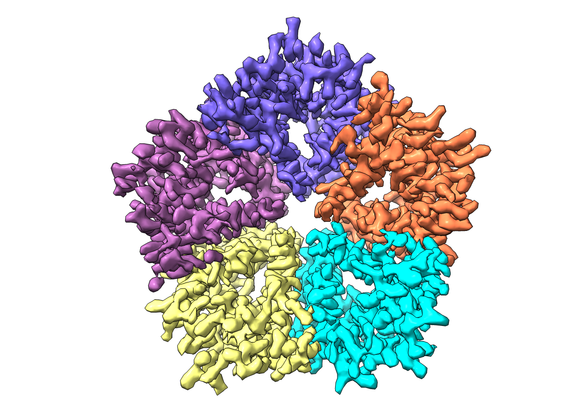 emd_21354.png emd_21354.png | 223.7 KB | ||
| Filedesc metadata | emd-21354.cif.gz | 4.9 KB | ||
| Archive directory |  http://ftp.pdbj.org/pub/emdb/structures/EMD-21354ftp://ftp.pdbj.org/pub/emdb/structures/EMD-21354 http://ftp.pdbj.org/pub/emdb/structures/EMD-21354ftp://ftp.pdbj.org/pub/emdb/structures/EMD-21354 | HTTPS FTP |
-Related structure data
| Related structure data |  6vqqMC  6vqrC  7mxyC M: atomic model generated by this map C: citing same article ( |
|---|---|
| Similar structure data |


-Links
| EMDB pages | EMDB (EBI/PDBe) / EMDataResource |
|---|---|
| Related items in Molecule of the Month |

-Map
| File | Download / File: emd_21354.map.gz / Format: CCP4 / Size: 2.7 MB / Type: IMAGE STORED AS FLOATING POINT NUMBER (4 BYTES) | ||||||||||||||||||||||||||||||||||||||||||||||||||||||||||||||||||||
|---|---|---|---|---|---|---|---|---|---|---|---|---|---|---|---|---|---|---|---|---|---|---|---|---|---|---|---|---|---|---|---|---|---|---|---|---|---|---|---|---|---|---|---|---|---|---|---|---|---|---|---|---|---|---|---|---|---|---|---|---|---|---|---|---|---|---|---|---|---|
| Voxel size | X=Y=Z: 1.08 Å | ||||||||||||||||||||||||||||||||||||||||||||||||||||||||||||||||||||
| Density |
| ||||||||||||||||||||||||||||||||||||||||||||||||||||||||||||||||||||
| Symmetry | Space group: 1 | ||||||||||||||||||||||||||||||||||||||||||||||||||||||||||||||||||||
| Details | EMDB XML:
CCP4 map header:
| ||||||||||||||||||||||||||||||||||||||||||||||||||||||||||||||||||||
-Supplemental data
- Sample components
-Entire : PfFNT
| Entire | Name: PfFNT |
|---|---|
| Components |
|
-Supramolecule #1: PfFNT
| Supramolecule | Name: PfFNT / type: complex / ID: 1 / Parent: 0 / Macromolecule list: all |
|---|---|
| Source (natural) | Organism: Plasmodium falciparum 3D7 (eukaryote) |
-Macromolecule #1: Formate-nitrite transporter
| Macromolecule | Name: Formate-nitrite transporter / type: protein_or_peptide / ID: 1 / Number of copies: 5 / Enantiomer: LEVO |
|---|---|
| Source (natural) | Organism: Plasmodium falciparum (isolate 3D7) (eukaryote) / Strain: isolate 3D7 |
| Molecular weight | Theoretical: 34.492281 KDa |
| Recombinant expression | Organism:  Homo sapiens (human) Homo sapiens (human) |
| Sequence | String: MPPNNSKYVL DPVSIKSVCG GEESYIRCVE YGKKKAHYSN LNLLAKAILA GMFVGLCAHA SGIAGGLFYY HKLREIVGAS MSVFVYGFT FPIAFMCIIC TGSDLFTGNT LAVTMALYEK KVKLLDYLRV MTISLFGNYV GAVSFAFFVS YLSGAFTNVH A VEKNHFFQ ...String: MPPNNSKYVL DPVSIKSVCG GEESYIRCVE YGKKKAHYSN LNLLAKAILA GMFVGLCAHA SGIAGGLFYY HKLREIVGAS MSVFVYGFT FPIAFMCIIC TGSDLFTGNT LAVTMALYEK KVKLLDYLRV MTISLFGNYV GAVSFAFFVS YLSGAFTNVH A VEKNHFFQ FLNDIAEKKV HHTFVECVSL AVGCNIFVCL AVYFVLTLKD GAGYVFSVFF AVYAFAIAGY EHIIANIYTL NI ALMVNTK ITVYQAYIKN LLPTLLGNYI AGAIVLGLPL YFIYKEHYYN FERSKRDNND AQMKSLSIEL RN UniProtKB: Formate-nitrite transporter |
-Experimental details
-Structure determination
| Method | cryo EM |
|---|---|
 Processing Processing | single particle reconstruction |
| Aggregation state | particle |
-Sample preparation
| Concentration | 0.5 mg/mL |
|---|---|
| Buffer | pH: 7.5 |
| Grid | Model: Quantifoil R1.2/1.3 / Material: COPPER / Mesh: 400 |
| Vitrification | Cryogen name: ETHANE / Chamber humidity: 100 % / Chamber temperature: 277 K / Instrument: FEI VITROBOT MARK IV |
| Details | This sample was monodisperse. |
- Electron microscopy
| Microscope | FEI TITAN KRIOS |
|---|---|
| Electron beam | Acceleration voltage: 300 kV / Electron source: FIELD EMISSION GUN |
| Electron optics | Illumination mode: SPOT SCAN / Imaging mode: BRIGHT FIELDBright-field microscopy |
| Image recording | Film or detector model: GATAN K3 BIOQUANTUM (6k x 4k) / Average electron dose: 29.0 e/Å2 |
| Experimental equipment |  Model: Titan Krios / Image courtesy: FEI Company |
-Image processing
| Startup model | Type of model: EMDB MAP |
|---|---|
| Initial angle assignment | Type: ANGULAR RECONSTITUTION |
| Final angle assignment | Type: ANGULAR RECONSTITUTION |
| Final reconstruction | Resolution.type: BY AUTHOR / Resolution: 2.56 Å / Resolution method: FSC 0.143 CUT-OFF / Number images used: 85976 |
