ムービー
ムービー コントローラー


+ データを開く

- 基本情報
基本情報
| 登録情報 | データベース: EMDB / ID: EMD-20308 | ||||||||||||
|---|---|---|---|---|---|---|---|---|---|---|---|---|---|
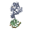
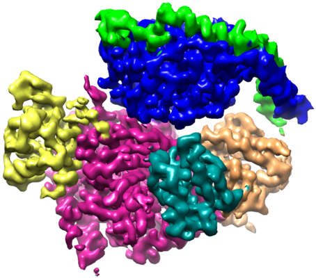
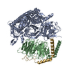
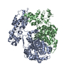
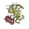
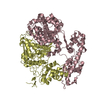
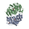
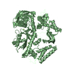
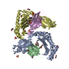
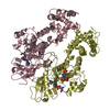
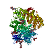
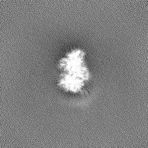
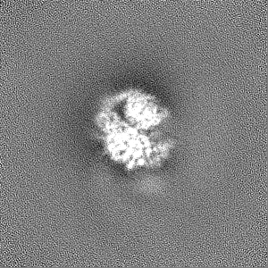
| タイトル | Single Particle Reconstruction of Phosphatidylinositol (3,4,5) trisphosphate-dependent Rac exchanger 1 bound to G protein beta gamma subunits | ||||||||||||
 マップデータ マップデータ | |||||||||||||
 試料 試料 |
| ||||||||||||
 キーワード キーワード | RhoGEF /  G protein (Gタンパク質) / Complex / Phosphatase fold / SIGNALING PROTEIN G protein (Gタンパク質) / Complex / Phosphatase fold / SIGNALING PROTEIN | ||||||||||||
| 機能・相同性 |  機能・相同性情報regulation of signaling / Olfactory Signaling Pathway / regulation of dendrite development / Sensory perception of sweet, bitter, and umami (glutamate) taste / Synthesis, secretion, and inactivation of Glucagon-like Peptide-1 (GLP-1) / regulation of actin filament polymerization / Activation of the phototransduction cascade / 好中球 / Activation of G protein gated Potassium channels / G-protein activation ...regulation of signaling / Olfactory Signaling Pathway / regulation of dendrite development / Sensory perception of sweet, bitter, and umami (glutamate) taste / Synthesis, secretion, and inactivation of Glucagon-like Peptide-1 (GLP-1) / regulation of actin filament polymerization / Activation of the phototransduction cascade / 好中球 / Activation of G protein gated Potassium channels / G-protein activation / G beta:gamma signalling through PI3Kgamma / Prostacyclin signalling through prostacyclin receptor / G beta:gamma signalling through PLC beta / ADP signalling through P2Y purinoceptor 1 / Thromboxane signalling through TP receptor / Presynaptic function of Kainate receptors / G beta:gamma signalling through CDC42 / Inhibition of voltage gated Ca2+ channels via Gbeta/gamma subunits / Glucagon-type ligand receptors / Adrenaline,noradrenaline inhibits insulin secretion / G alpha (12/13) signalling events / G beta:gamma signalling through BTK / ADP signalling through P2Y purinoceptor 12 / Cooperation of PDCL (PhLP1) and TRiC/CCT in G-protein beta folding / regulation of small GTPase mediated signal transduction / Thrombin signalling through proteinase activated receptors (PARs) / Ca2+ pathway / G alpha (z) signalling events / Extra-nuclear estrogen signaling / G alpha (s) signalling events / G alpha (q) signalling events / G alpha (i) signalling events / Glucagon-like Peptide-1 (GLP1) regulates insulin secretion / Vasopressin regulates renal water homeostasis via Aquaporins / RHOB GTPase cycle / NRAGE signals death through JNK / superoxide metabolic process / RHOJ GTPase cycle / RHOC GTPase cycle / RHOQ GTPase cycle / CDC42 GTPase cycle / RHOG GTPase cycle / T cell differentiation / RHOA GTPase cycle / RAC2 GTPase cycle / RAC3 GTPase cycle / positive regulation of substrate adhesion-dependent cell spreading / RAC1 GTPase cycle / actin filament polymerization / GTPase activator activity / 好中球 / guanyl-nucleotide exchange factor activity / dendritic shaft / phospholipid binding / photoreceptor disc membrane / cellular response to catecholamine stimulus / adenylate cyclase-activating dopamine receptor signaling pathway / cellular response to prostaglandin E stimulus / G-protein beta-subunit binding / heterotrimeric G-protein complex / G alpha (12/13) signalling events / signaling receptor complex adaptor activity / 成長円錐 / intracellular signal transduction / positive regulation of cell migration / G protein-coupled receptor signaling pathway / GTPase activity / perinuclear region of cytoplasm / enzyme binding / 生体膜 / 細胞膜 / 細胞質基質 / 細胞質 機能・相同性情報regulation of signaling / Olfactory Signaling Pathway / regulation of dendrite development / Sensory perception of sweet, bitter, and umami (glutamate) taste / Synthesis, secretion, and inactivation of Glucagon-like Peptide-1 (GLP-1) / regulation of actin filament polymerization / Activation of the phototransduction cascade / 好中球 / Activation of G protein gated Potassium channels / G-protein activation ...regulation of signaling / Olfactory Signaling Pathway / regulation of dendrite development / Sensory perception of sweet, bitter, and umami (glutamate) taste / Synthesis, secretion, and inactivation of Glucagon-like Peptide-1 (GLP-1) / regulation of actin filament polymerization / Activation of the phototransduction cascade / 好中球 / Activation of G protein gated Potassium channels / G-protein activation / G beta:gamma signalling through PI3Kgamma / Prostacyclin signalling through prostacyclin receptor / G beta:gamma signalling through PLC beta / ADP signalling through P2Y purinoceptor 1 / Thromboxane signalling through TP receptor / Presynaptic function of Kainate receptors / G beta:gamma signalling through CDC42 / Inhibition of voltage gated Ca2+ channels via Gbeta/gamma subunits / Glucagon-type ligand receptors / Adrenaline,noradrenaline inhibits insulin secretion / G alpha (12/13) signalling events / G beta:gamma signalling through BTK / ADP signalling through P2Y purinoceptor 12 / Cooperation of PDCL (PhLP1) and TRiC/CCT in G-protein beta folding / regulation of small GTPase mediated signal transduction / Thrombin signalling through proteinase activated receptors (PARs) / Ca2+ pathway / G alpha (z) signalling events / Extra-nuclear estrogen signaling / G alpha (s) signalling events / G alpha (q) signalling events / G alpha (i) signalling events / Glucagon-like Peptide-1 (GLP1) regulates insulin secretion / Vasopressin regulates renal water homeostasis via Aquaporins / RHOB GTPase cycle / NRAGE signals death through JNK / superoxide metabolic process / RHOJ GTPase cycle / RHOC GTPase cycle / RHOQ GTPase cycle / CDC42 GTPase cycle / RHOG GTPase cycle / T cell differentiation / RHOA GTPase cycle / RAC2 GTPase cycle / RAC3 GTPase cycle / positive regulation of substrate adhesion-dependent cell spreading / RAC1 GTPase cycle / actin filament polymerization / GTPase activator activity / 好中球 / guanyl-nucleotide exchange factor activity / dendritic shaft / phospholipid binding / photoreceptor disc membrane / cellular response to catecholamine stimulus / adenylate cyclase-activating dopamine receptor signaling pathway / cellular response to prostaglandin E stimulus / G-protein beta-subunit binding / heterotrimeric G-protein complex / G alpha (12/13) signalling events / signaling receptor complex adaptor activity / 成長円錐 / intracellular signal transduction / positive regulation of cell migration / G protein-coupled receptor signaling pathway / GTPase activity / perinuclear region of cytoplasm / enzyme binding / 生体膜 / 細胞膜 / 細胞質基質 / 細胞質類似検索 - 分子機能 | ||||||||||||
| 生物種 |  Homo sapiens (ヒト) / Homo sapiens (ヒト) /  Bos taurus (ウシ) Bos taurus (ウシ) | ||||||||||||
| 手法 | 単粒子再構成法 / クライオ電子顕微鏡法 / 解像度: 3.2 Å | ||||||||||||
 データ登録者 データ登録者 | Cash JN / Cianfrocco MA | ||||||||||||
| 資金援助 |  米国, 3件 米国, 3件
| ||||||||||||
 引用 引用 | ジャーナル: Sci Adv / 年: 2019 タイトル: Cryo-electron microscopy structure and analysis of the P-Rex1-Gβγ signaling scaffold. 著者: Jennifer N Cash / Sarah Urata / Sheng Li / Sandeep K Ravala / Larisa V Avramova / Michael D Shost / J Silvio Gutkind / John J G Tesmer / Michael A Cianfrocco / 要旨: PIP-dependent Rac exchanger 1 (P-Rex1) is activated downstream of G protein-coupled receptors to promote neutrophil migration and metastasis. The structure of more than half of the enzyme and its ...PIP-dependent Rac exchanger 1 (P-Rex1) is activated downstream of G protein-coupled receptors to promote neutrophil migration and metastasis. The structure of more than half of the enzyme and its regulatory G protein binding site are unknown. Our 3.2 Å cryo-EM structure of the P-Rex1-Gβγ complex reveals that the carboxyl-terminal half of P-Rex1 adopts a complex fold most similar to those of phosphoinositide phosphatases. Although catalytically inert, the domain coalesces with a DEP domain and two PDZ domains to form an extensive docking site for Gβγ. Hydrogen-deuterium exchange mass spectrometry suggests that Gβγ binding induces allosteric changes in P-Rex1, but functional assays indicate that membrane localization is also required for full activation. Thus, a multidomain assembly is key to the regulation of P-Rex1 by Gβγ and the formation of a membrane-localized scaffold optimized for recruitment of other signaling proteins such as PKA and PTEN. | ||||||||||||
| 履歴 |
|
- 構造の表示
構造の表示
| ムービー |
ムービービューア |
|---|---|
| 構造ビューア | EMマップ: SurfViewMolmilJmol/JSmol |
| 添付画像 |
- ダウンロードとリンク
-EMDBアーカイブ
| マップデータ | emd_20308.map.gz | 52.5 MB | EMDBマップデータ形式 | |
|---|---|---|---|---|
| ヘッダ (付随情報) | emd-20308-v30.xmlemd-20308.xml | 27.5 KB 27.5 KB | 表示 表示 | EMDBヘッダ |
| FSC (解像度算出) | emd_20308_fsc.xml | 12.5 KB | 表示 | FSCデータファイル |
| 画像 |  emd_20308.png emd_20308.png | 215.7 KB | ||
| マスクデータ | emd_20308_msk_1.map | 103 MB | マスクマップ | |
| Filedesc metadata | emd-20308.cif.gz | 7.5 KB | ||
| その他 | emd_20308_additional_1.map.gzemd_20308_additional_2.map.gzemd_20308_half_map_1.map.gzemd_20308_half_map_2.map.gz | 97.2 MB 51.9 MB 95.5 MB 95.5 MB | ||
| アーカイブディレクトリ |  http://ftp.pdbj.org/pub/emdb/structures/EMD-20308ftp://ftp.pdbj.org/pub/emdb/structures/EMD-20308 http://ftp.pdbj.org/pub/emdb/structures/EMD-20308ftp://ftp.pdbj.org/pub/emdb/structures/EMD-20308 | HTTPS FTP |
-関連構造データ
| 関連構造データ |  6pcvMC M: このマップから作成された原子モデル C: 同じ文献を引用 ( |
|---|---|
| 類似構造データ | |
| 電子顕微鏡画像生データ | EMPIAR-10285 (タイトル: Cryo-electron microscopy structure of the P-Rex1–G-beta-gamma signaling scaffold Data size: 3.0 TB Data #1: Movie files (.tif) for P-Rex1-Gbg [micrographs - multiframe] Data #2: Micrograph files (.mrc) & CTF log files for P-Rex1-Gbg [micrographs - single frame] Data #3: Extracted particles from Warp for P-Rex1-Gbg [picked particles - single frame - processed]) |

-リンク
| EMDBのページ | EMDB (EBI/PDBe) / EMDataResource |
|---|---|
| 「今月の分子」の関連する項目 |


-マップ
| ファイル | ダウンロード / ファイル: emd_20308.map.gz / 形式: CCP4 / 大きさ: 103 MB / タイプ: IMAGE STORED AS FLOATING POINT NUMBER (4 BYTES) | ||||||||||||||||||||||||||||||||||||||||||||||||||||||||||||||||||||
|---|---|---|---|---|---|---|---|---|---|---|---|---|---|---|---|---|---|---|---|---|---|---|---|---|---|---|---|---|---|---|---|---|---|---|---|---|---|---|---|---|---|---|---|---|---|---|---|---|---|---|---|---|---|---|---|---|---|---|---|---|---|---|---|---|---|---|---|---|---|
| ボクセルのサイズ | X=Y=Z: 1 Å | ||||||||||||||||||||||||||||||||||||||||||||||||||||||||||||||||||||
| 密度 |
| ||||||||||||||||||||||||||||||||||||||||||||||||||||||||||||||||||||
| 対称性 | 空間群: 1 | ||||||||||||||||||||||||||||||||||||||||||||||||||||||||||||||||||||
| 詳細 | EMDB XML:
CCP4マップ ヘッダ情報:
| ||||||||||||||||||||||||||||||||||||||||||||||||||||||||||||||||||||
-添付データ
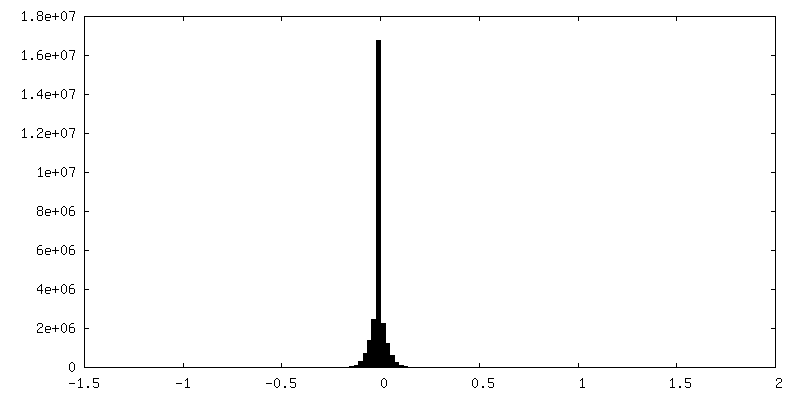
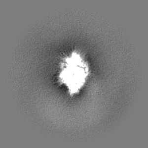
-マスク #1
| ファイル | emd_20308_msk_1.map | ||||||||||||
|---|---|---|---|---|---|---|---|---|---|---|---|---|---|
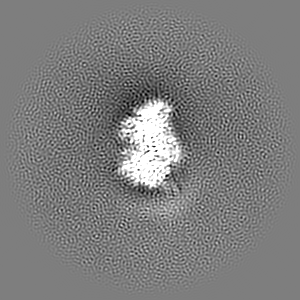
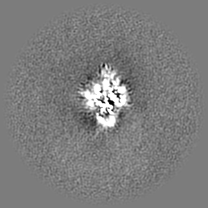
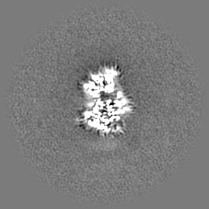
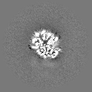
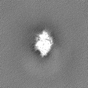
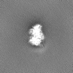
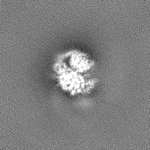
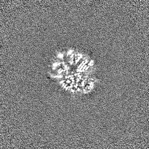
| 投影像・断面図 |
| ||||||||||||
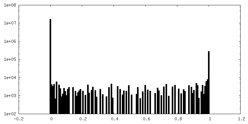
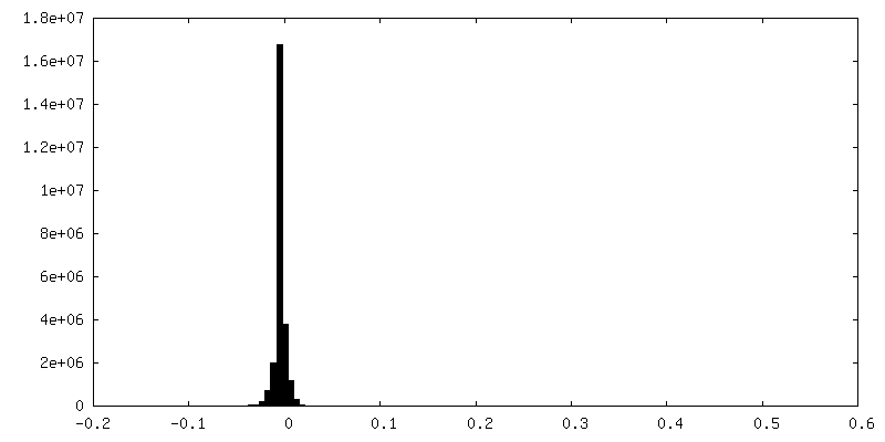
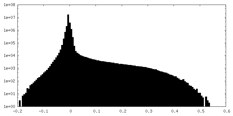
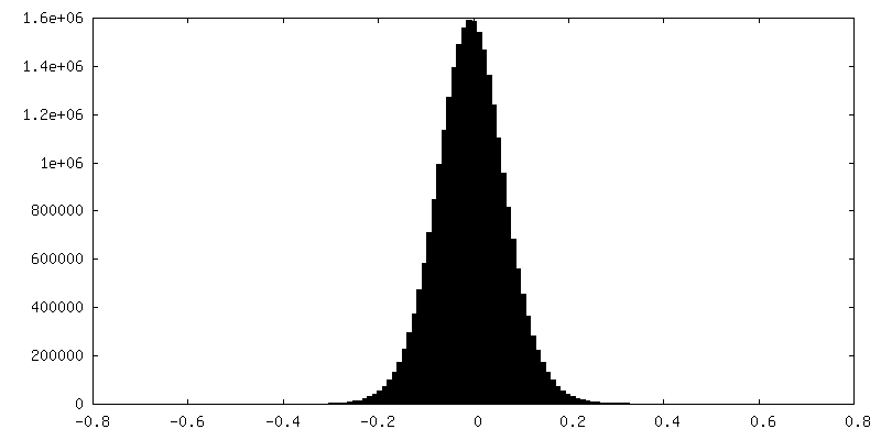
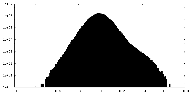
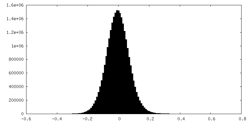
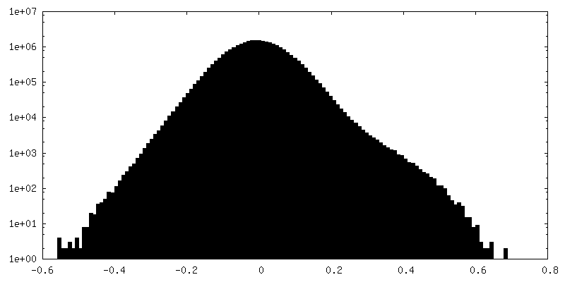
| 密度ヒストグラム |
 Z
Z Y
Y X
X


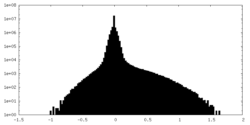
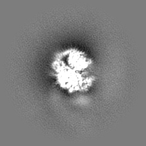
-追加マップ: Sharpened map
| ファイル | emd_20308_additional_1.map | ||||||||||||
|---|---|---|---|---|---|---|---|---|---|---|---|---|---|
| 注釈 | Sharpened map | ||||||||||||
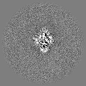
| 投影像・断面図 |
| ||||||||||||
| 密度ヒストグラム |


-追加マップ: Unsharpened map
| ファイル | emd_20308_additional_2.map | ||||||||||||
|---|---|---|---|---|---|---|---|---|---|---|---|---|---|
| 注釈 | Unsharpened map | ||||||||||||
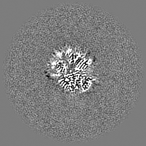
| 投影像・断面図 |
| ||||||||||||
| 密度ヒストグラム |

-ハーフマップ: #2
| ファイル | emd_20308_half_map_1.map | ||||||||||||
|---|---|---|---|---|---|---|---|---|---|---|---|---|---|
| 投影像・断面図 |
| ||||||||||||
| 密度ヒストグラム |

-ハーフマップ: #1
| ファイル | emd_20308_half_map_2.map | ||||||||||||
|---|---|---|---|---|---|---|---|---|---|---|---|---|---|
| 投影像・断面図 |
| ||||||||||||
| 密度ヒストグラム |

- 試料の構成要素
-全体 : P-Rex1 bound to G protein beta gamma subunits
| 全体 | 名称: P-Rex1 bound to G protein beta gamma subunits |
|---|---|
| 要素 |
|
-超分子 #1: P-Rex1 bound to G protein beta gamma subunits
| 超分子 | 名称: P-Rex1 bound to G protein beta gamma subunits / タイプ: complex / ID: 1 / 親要素: 0 / 含まれる分子: all |
|---|---|
| 由来(天然) | 生物種: Homo sapiens (ヒト) |
| 分子量 | 理論値: 9 KDa |
-超分子 #2: Phosphatidylinositol (3,4,5) trisphosphate-dependent Rac exchanger 1
| 超分子 | 名称: Phosphatidylinositol (3,4,5) trisphosphate-dependent Rac exchanger 1 タイプ: complex / ID: 2 / 親要素: 1 / 含まれる分子: #1 |
|---|
-超分子 #3: G protein beta-1 subunit
| 超分子 | 名称: G protein beta-1 subunit / タイプ: complex / ID: 3 / 親要素: 1 / 含まれる分子: #2 |
|---|
-超分子 #4: G protein gamma-2 subunit
| 超分子 | 名称: G protein gamma-2 subunit / タイプ: complex / ID: 4 / 親要素: 1 / 含まれる分子: #3 |
|---|
-分子 #1: Phosphatidylinositol (3,4,5) trisphosphate-dependent Rac exchanger 1
| 分子 | 名称: Phosphatidylinositol (3,4,5) trisphosphate-dependent Rac exchanger 1 タイプ: protein_or_peptide / ID: 1 / コピー数: 1 / 光学異性体: LEVO |
|---|---|
| 由来(天然) | 生物種: Homo sapiens (ヒト) |
| 分子量 | 理論値: 184.840891 KDa |
| 組換発現 | 生物種: Homo sapiens (ヒト) |
| 配列 | 文字列: GEFAAARESE RQLRLRLCVL NEILGTERDY VGTLRFLQSA FLHRIRQNVA DSVEKGLTEE NVKVLFSNIE DILEVHKDFL AALEYCLHP EPQSQHELGN VFLKFKDKFC VYEEYCSNHE KALRLLVELN KIPTVRAFLL SCMLLGGRKT TDIPLEGYLL S PIQRICKY ...文字列: GEFAAARESE RQLRLRLCVL NEILGTERDY VGTLRFLQSA FLHRIRQNVA DSVEKGLTEE NVKVLFSNIE DILEVHKDFL AALEYCLHP EPQSQHELGN VFLKFKDKFC VYEEYCSNHE KALRLLVELN KIPTVRAFLL SCMLLGGRKT TDIPLEGYLL S PIQRICKY PLLLKELAKR TPGKHPDHPA VQSALQAMKT VCSNINETKR QMEKLEALEQ LQSHIEGWEG SNLTDICTQL LL QGTLLKI SAGNIQERAF FLFDNLLVYC KRKSRVTGSK KSTKRTKSIN GSLYIFRGRI NTEVMEVENV EDGTADYHSN GYT VTNGWK IHNTAKNKWF VCMAKTAEEK QKWLDAIIRE REQRESLKLG MERDAYVMIA EKGEKLYHMM MNKKVNLIKD RRRK LSTVP KCFLGNEFVA WLLEIGEISK TEEGVNLGQA LLENGIIHHV SDKHQFKNEQ VMYRFRYDDG TYKARSELED IMSKG VRLY CRLHSLYTPV IKDRDYHLKT YKSVLPGSKL VDWLLAQGDC QTREEAVALG VGLCNNGFMH HVLEKSEFRD ESQYFR FHA DEEMEGTSSK NKQLRNDFKL VENILAKRLL ILPQEEDYGF DIEEKNKAVV VKSVQRGSLA EVAGLQVGRK IYSINED LV FLRPFSEVES ILNQSFCSRR PLRLLVATKA KEIIKIPDQP DTLCFQIRGA APPYVYAVGR GSEAMAAGLC AGQCILKV N GSNVMNDGAP EVLEHFQAFR SRREEALGLY QWIYHTHEDA QEARASQEAS TEDPSGEQAQ EEDQADSAFP LLSLGPRLS LCEDSPMVTL TVDNVHLEHG VVYEYVSTAG VRCHVLEKIV EPRGCFGLTA KILEAFAAND SVFVENCRRL MALSSAIVTM PHFEFRNIC DTKLESIGQR IACYQEFAAQ LKSRVSPPFK QAPLEPHPLC GLDFCPTNCH INLMEVSYPK TTPSVGRSFS I RFGRKPSL IGLDPEQGHL NPMSYTQHCI TTMAAPSWKC LPAAEGDPQG QGLHDGSFGP ASGTLGQEDR GLSFLLKQED RE IQDAYLQ LFTKLDVALK EMKQYVTQIN RLLSTITEPT SGGSCDASLA EEASSLPLVS EESEMDRSDH GGIKKVCFKV AEE DQEDSG HDTMSYRDSY SECNSNRDSV LSYTSVRSNS SYLGSDEMGS GDELPCDMRI PSDKQDKLHG CLEHLFNQVD SINA LLKGP VMSRAFEETK HFPMNHSLQE FKQKEECTIR GRSLIQISIQ EDPWNLPNSI KTLVDNIQRY VEDGKNQLLL ALLKC TDTE LQLRRDAIFC QALVAAVCTF SEQLLAALGY RYNNNGEYEE SSRDASRKWL EQVAATGVLL HCQSLLSPAT VKEERT MLE DIWVTLSELD NVTFSFKQLD ENYVANTNVF YHIEGSRQAL KVIFYLDSYH FSKLPSRLEG GASLRLHTAL FTKVLEN VE GLPSPGSQAA EDLQQDINAQ SLEKVQQYYR KLRAFYLERS NLPTDASTTA VKIDQLIRPI NALDELCRLM KSFVHPKP G AAGSVGAGLI PISSELCYRL GACQMVMCGT GMQRSTLSVS LEQAAILARS HGLLPKCIMQ ATDIMRKQGP RVEILAKNL RVKDQMPQGA PRLYRLCQPP VDGDLHHHHH HHHHH UniProtKB: PREX1 |
-分子 #2: Guanine nucleotide-binding protein G(I)/G(S)/G(T) subunit beta-1
| 分子 | 名称: Guanine nucleotide-binding protein G(I)/G(S)/G(T) subunit beta-1 タイプ: protein_or_peptide / ID: 2 / コピー数: 1 / 光学異性体: LEVO |
|---|---|
| 由来(天然) | 生物種: Bos taurus (ウシ) |
| 分子量 | 理論値: 37.41693 KDa |
| 組換発現 | 生物種:  Trichoplusia ni (イラクサキンウワバ) Trichoplusia ni (イラクサキンウワバ) |
| 配列 | 文字列: MSELDQLRQE AEQLKNQIRD ARKACADATL SQITNNIDPV GRIQMRTRRT LRGHLAKIYA MHWGTDSRLL VSASQDGKLI IWDSYTTNK VHAIPLRSSW VMTCAYAPSG NYVACGGLDN ICSIYNLKTR EGNVRVSREL AGHTGYLSCC RFLDDNQIVT S SGDTTCAL ...文字列: MSELDQLRQE AEQLKNQIRD ARKACADATL SQITNNIDPV GRIQMRTRRT LRGHLAKIYA MHWGTDSRLL VSASQDGKLI IWDSYTTNK VHAIPLRSSW VMTCAYAPSG NYVACGGLDN ICSIYNLKTR EGNVRVSREL AGHTGYLSCC RFLDDNQIVT S SGDTTCAL WDIETGQQTT TFTGHTGDVM SLSLAPDTRL FVSGACDASA KLWDVREGMC RQTFTGHESD INAICFFPNG NA FATGSDD ATCRLFDLRA DQELMTYSHD NIICGITSVS FSKSGRLLLA GYDDFNCNVW DALKADRAGV LAGHDNRVSC LGV TDDGMA VATGSWDSFL KIWN UniProtKB: Guanine nucleotide-binding protein G(I)/G(S)/G(T) subunit beta-1 |
-分子 #3: Guanine nucleotide-binding protein G(I)/G(S)/G(O) subunit gamma-2
| 分子 | 名称: Guanine nucleotide-binding protein G(I)/G(S)/G(O) subunit gamma-2 タイプ: protein_or_peptide / ID: 3 / コピー数: 1 / 光学異性体: LEVO |
|---|---|
| 由来(天然) | 生物種: Bos taurus (ウシ) |
| 分子量 | 理論値: 9.226547 KDa |
| 組換発現 | 生物種: Trichoplusia ni (イラクサキンウワバ) |
| 配列 | 文字列: HHHHHHHHHH MASNNTASIA QARKLVEQLK MEANIDRIKV SKAAADLMAY CEAHAKEDPL LTPVPASENP FREKKFFSAI L UniProtKB: Guanine nucleotide-binding protein G(I)/G(S)/G(O) subunit gamma-2 |
-実験情報
-構造解析
| 手法 | クライオ電子顕微鏡法 |
|---|---|
 解析 解析 | 単粒子再構成法 |
| 試料の集合状態 | particle |
-試料調製
| 濃度 | 0.7 mg/mL |
|---|---|
| 緩衝液 | pH: 8 |
| グリッド | モデル: Quantifoil, UltrAuFoil, R1.2/1.3 / 材質: GOLD / メッシュ: 300 / 前処理 - タイプ: GLOW DISCHARGE / 前処理 - 時間: 75 sec. |
| 凍結 | 凍結剤: ETHANE / チャンバー内湿度: 100 % / チャンバー内温度: 277 K / 装置: FEI VITROBOT MARK IV |
- 電子顕微鏡法
| 顕微鏡 | FEI TITAN KRIOS |
|---|---|
| 電子線 | 加速電圧: 300 kV / 電子線源: FIELD EMISSION GUN |
| 電子光学系 | C2レンズ絞り径: 70.0 µm / 照射モード: OTHER / 撮影モード: BRIGHT FIELDBright-field microscopy / Cs: 2.7 mm / 倍率(公称値): 29000 |
| 試料ステージ | 試料ホルダーモデル: FEI TITAN KRIOS AUTOGRID HOLDER ホルダー冷却材: NITROGEN |
| 撮影 | フィルム・検出器のモデル: GATAN K2 SUMMIT (4k x 4k) 検出モード: COUNTING / デジタル化 - サイズ - 横: 3838 pixel / デジタル化 - サイズ - 縦: 3710 pixel / 撮影したグリッド数: 2 / 実像数: 6746 / 平均電子線量: 47.0 e/Å2 |
| 実験機器 |  モデル: Titan Krios / 画像提供: FEI Company |
-画像解析
| 粒子像選択 | 選択した数: 905464 詳細: 600,588 particles (untilted) and 304,876 particles (30 degree tilted) |
|---|---|
| 初期モデル | モデルのタイプ: OTHER 詳細: cryoSPARC ab initio model calculation was used to create initial model |
| 初期 角度割当 | タイプ: MAXIMUM LIKELIHOOD / ソフトウェア - 名称: cryoSPARC (ver. v0.65) |
| 最終 角度割当 | タイプ: MAXIMUM LIKELIHOOD / ソフトウェア - 名称: cryoSPARC (ver. v0.65) |
| 最終 再構成 | 想定した対称性 - 点群: C1 (非対称) / 解像度のタイプ: BY AUTHOR / 解像度: 3.2 Å / 解像度の算出法: FSC 0.143 CUT-OFF / ソフトウェア - 名称: cryoSPARC (ver. v0.65) / 使用した粒子像数: 205599 |
| FSC曲線 (解像度の算出) | 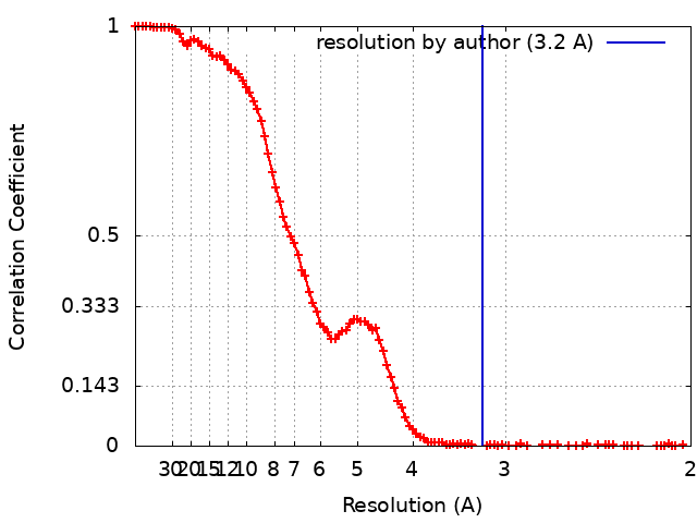 |
-原子モデル構築 1
| 初期モデル |
| ||||||||
|---|---|---|---|---|---|---|---|---|---|
| 精密化 | 空間: REAL / プロトコル: AB INITIO MODEL / 温度因子: 83 | ||||||||
| 得られたモデル | PDB-6pcv: |