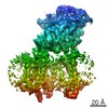
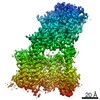
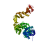
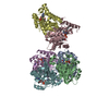
登録情報 データベース : EMDB / ID : EMD-20222タイトル Structure of the Rhodopsin-Transducin Complex Rhodopsin-Transducin Complex 複合体 : Rhodopsin-Transducin Complex複合体 : Transducinタンパク質・ペプチド : Gt-alpha/Gi1-alpha chimeraタンパク質・ペプチド : Guanine nucleotide-binding protein G(I)/G(S)/G(T) subunit beta-1タンパク質・ペプチド : Guanine nucleotide-binding protein G(T) subunit gamma-T1複合体 : Rhodopsinタンパク質・ペプチド : Rhodopsinリガンド : RETINAL機能・相同性 分子機能 ドメイン・相同性 構成要素
/ / / / / / / / / / / / / / / / / / / / / / / / / / / / / / / / / / / / / / / / / / / / / / / / / / / / / / / / / / / / / / / / / / / / / / / / / / / / / / / / / / / / / / / / / / / / / / / / / / / / / / / / / / / / / / / / / / / / / / / / / / / / / / / / / / / / / / / / / / / / / / / / / / 生物種 Bos taurus (ウシ) / Bovine (ウシ)手法 / / 解像度 : 3.9 Å Gao Y / Hu H / Ramachandran S / Erickson JW / Cerione RA / Skiniotis G 資金援助 Organization Grant number 国 National Institutes of Health/National Cancer Institute (NIH/NCI) R01 CA201402 National Institutes of Health/National Institute of Neurological Disorders and Stroke (NIH/NINDS) R01 NS092695
ジャーナル : Mol Cell / 年 : 2019タイトル : Structures of the Rhodopsin-Transducin Complex: Insights into G-Protein Activation.著者 : Yang Gao / Hongli Hu / Sekar Ramachandran / Jon W Erickson / Richard A Cerione / Georgios Skiniotis / 要旨 : Rhodopsin (Rho), a prototypical G-protein-coupled receptor (GPCR) in vertebrate vision, activates the G-protein transducin (G) by catalyzing GDP-GTP exchange on its α subunit (Gα). To elucidate the ... Rhodopsin (Rho), a prototypical G-protein-coupled receptor (GPCR) in vertebrate vision, activates the G-protein transducin (G) by catalyzing GDP-GTP exchange on its α subunit (Gα). To elucidate the determinants of G coupling and activation, we obtained cryo-EM structures of a fully functional, light-activated Rho-G complex in the presence and absence of a G-protein-stabilizing nanobody. The structures illustrate how G overcomes its low basal activity by engaging activated Rho in a conformation distinct from other GPCR-G-protein complexes. Moreover, the nanobody-free structures reveal native conformations of G-protein components and capture three distinct conformers showing the Gα helical domain (αHD) contacting the Gβγ subunits. These findings uncover the molecular underpinnings of G-protein activation by visual rhodopsin and shed new light on the role played by Gβγ during receptor-catalyzed nucleotide exchange. 履歴 登録 2019年5月14日 - ヘッダ(付随情報) 公開 2019年6月19日 - マップ公開 2019年7月24日 - 更新 2019年12月4日 - 現状 2019年12月4日 処理サイト : RCSB / 状態 : 公開
すべて表示 表示を減らす
 ムービー
ムービー コントローラー


 データを開く
 基本情報
基本情報 マップデータ
 試料
試料 機能・相同性情報
 Opsins / VxPx cargo-targeting to cilium / rod bipolar cell differentiation / rod photoreceptor outer segment / sperm head plasma membrane / Olfactory Signaling Pathway / absorption of visible light / Sensory perception of sweet, bitter, and umami (glutamate) taste ...detection of light stimulus involved in visual perception / negative regulation of cyclic-nucleotide phosphodiesterase activity /
 データ登録者
 米国, 2件
  引用
引用 構造の表示
構造の表示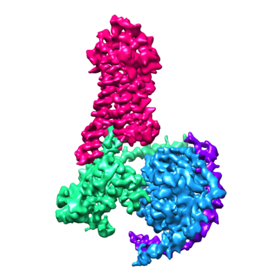
 ダウンロードとリンク
 emd_20222.png
 http://ftp.pdbj.org/pub/emdb/structures/EMD-20222


 試料の構成要素

 解析
解析 電子顕微鏡法