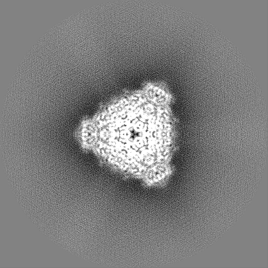
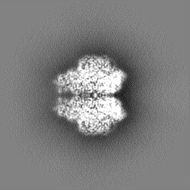
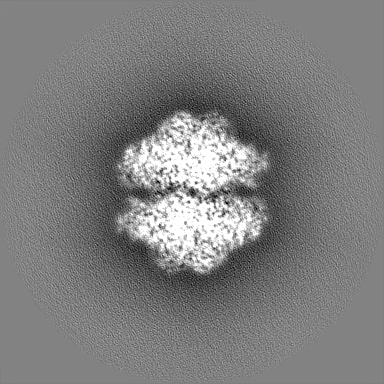
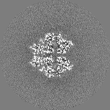
Journal: J Struct Biol X / Year: 2021 Title: Experimental evaluation of super-resolution imaging and magnification choice in single-particle cryo-EM. Authors: J Ryan Feathers / Katherine A Spoth / J Christopher Fromme / Abstract: The resolution of cryo-EM reconstructions is fundamentally limited by the Nyquist frequency, which is half the sampling frequency of the detector and depends upon the magnification used. In ...The resolution of cryo-EM reconstructions is fundamentally limited by the Nyquist frequency, which is half the sampling frequency of the detector and depends upon the magnification used. In principle, super-resolution imaging should enable reconstructions to surpass the physical Nyquist limit by increasing sampling frequency, yet there are few reports of reconstructions that do so. Here we directly examine the contribution of super-resolution information, obtained with the K3 direct electron detector using a 2-condenser microscope, to single-particle cryo-EM reconstructions surpassing the physical Nyquist limit. We also present a comparative analysis of a sample imaged at four different magnifications. This analysis demonstrates that lower magnifications can be beneficial, despite the loss of higher resolution signal, due to the increased number of particle images obtained. To highlight the potential utility of lower magnification data collection, we produced a 3.5 Å reconstruction of jack bean urease with particles from a single micrograph.
History
Deposition
Mar 22, 2019
-
Header (metadata) release
May 1, 2019
-
Map release
Mar 25, 2020
-
Update
Apr 21, 2021
-
Current status
Apr 21, 2021
Processing site: RCSB / Status: Released
-
Structure visualization
Movie
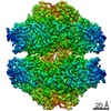
Surface view with section colored by density value
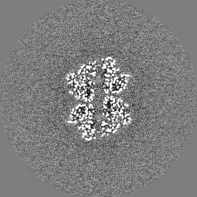
EMPIAR-10547 (Title: Jack bean urease imaged at 49kX nominal magnification Data size: 169.3 / Data #1: Super resolution movies [micrographs - multiframe])
In the structure databanks used in Yorodumi, some data are registered as the other names, "COVID-19 virus" and "2019-nCoV". Here are the details of the virus and the list of structure data.
Jan 31, 2019. EMDB accession codes are about to change! (news from PDBe EMDB page)
EMDB accession codes are about to change! (news from PDBe EMDB page)
The allocation of 4 digits for EMDB accession codes will soon come to an end. Whilst these codes will remain in use, new EMDB accession codes will include an additional digit and will expand incrementally as the available range of codes is exhausted. The current 4-digit format prefixed with “EMD-” (i.e. EMD-XXXX) will advance to a 5-digit format (i.e. EMD-XXXXX), and so on. It is currently estimated that the 4-digit codes will be depleted around Spring 2019, at which point the 5-digit format will come into force.
The EM Navigator/Yorodumi systems omit the EMD- prefix.
Related info.:Q: What is EMD? / ID/Accession-code notation in Yorodumi/EM Navigator
Yorodumi is a browser for structure data from EMDB, PDB, SASBDB, etc.
This page is also the successor to EM Navigator detail page, and also detail information page/front-end page for Omokage search.
The word "yorodu" (or yorozu) is an old Japanese word meaning "ten thousand". "mi" (miru) is to see.
Related info.:EMDB / PDB / SASBDB / Comparison of 3 databanks / Yorodumi Search / Aug 31, 2016. New EM Navigator & Yorodumi / Yorodumi Papers / Jmol/JSmol / Function and homology information / Changes in new EM Navigator and Yorodumi
 Movie
Movie Controller

 Yorodumi
 Open data
 Basic information
 Map data
 Sample
 Function and homology information
 urease complex /

 Authors
 United States, 1 items
  Citation
 Structure visualization
 Downloads & links
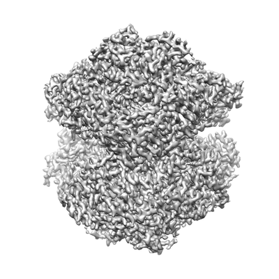 emd_20016.png
 http://ftp.pdbj.org/pub/emdb/structures/EMD-20016


 Z
Z Y
Y X
X


 Sample components
 Processing
 Electron microscopy


