 Movie
Movie Controller


+ Open data

- Basic information
| Entry |  | |||||||||
|---|---|---|---|---|---|---|---|---|---|---|
| Title | Trypanosoma brucei 3-methylcrotonyl-CoA carboxylase | |||||||||
 Map data Map data | volume map sharp | |||||||||
 Sample Sample |
| |||||||||
 Keywords Keywords |  carboxylase / trypanosoma brucei / BIOSYNTHETIC PROTEIN / TRANSFERASE carboxylase / trypanosoma brucei / BIOSYNTHETIC PROTEIN / TRANSFERASE | |||||||||
| Function / homology |  Function and homology informationmethylcrotonoyl-CoA carboxylase / methylcrotonoyl-CoA carboxylase activity / methylcrotonoyl-CoA carboxylase complex / L-leucine catabolic process / pyrimidine nucleobase biosynthetic process / biotin binding / cilium / mitochondrion / ATP binding / metal ion binding / cytoplasm Function and homology informationmethylcrotonoyl-CoA carboxylase / methylcrotonoyl-CoA carboxylase activity / methylcrotonoyl-CoA carboxylase complex / L-leucine catabolic process / pyrimidine nucleobase biosynthetic process / biotin binding / cilium / mitochondrion / ATP binding / metal ion binding / cytoplasmSimilarity search - Function | |||||||||
| Biological species |  Trypanosoma brucei (eukaryote) Trypanosoma brucei (eukaryote) | |||||||||
| Method | single particle reconstruction / cryo EM / Resolution: 2.37 Å | |||||||||
 Authors Authors | Ruiz FM / Plaza-Pegueroles A / Fernandez-Tornero C | |||||||||
| Funding support |  Spain, 1 items Spain, 1 items
| |||||||||
 Citation Citation | Journal: Structure / Year: 2024 Title: The cryo-EM structure of trypanosome 3-methylcrotonyl-CoA carboxylase provides mechanistic and dynamic insights into its enzymatic function. Authors: Adrián Plaza-Pegueroles / Inna Aphasizheva / Ruslan Aphasizhev / Carlos Fernández-Tornero / Federico M Ruiz /  Abstract: 3-Methylcrotonyl-CoA carboxylase (MCC) catalyzes the two-step, biotin-dependent production of 3-methylglutaconyl-CoA, an essential intermediate in leucine catabolism. Given the critical metabolic ...3-Methylcrotonyl-CoA carboxylase (MCC) catalyzes the two-step, biotin-dependent production of 3-methylglutaconyl-CoA, an essential intermediate in leucine catabolism. Given the critical metabolic role of MCC, deficiencies in this enzyme lead to organic aciduria, while its overexpression is linked to tumor development. MCC is a dodecameric enzyme composed of six copies of each α- and β-subunit. We present the cryo-EM structure of the endogenous MCC holoenzyme from Trypanosoma brucei in a non-filamentous state at 2.4 Å resolution. Biotin is covalently bound to the biotin carboxyl carrier protein domain of α-subunits and positioned in a non-canonical pocket near the active site of neighboring β-subunit dimers. Moreover, flexibility of key residues at α-subunit interfaces and loops enables pivoting of α-subunit trimers to partly reduce the distance between α- and β-subunit active sites, required for MCC catalysis. Our results provide a structural framework to understand the enzymatic mechanism of eukaryotic MCCs and to assist drug discovery against trypanosome infections. | |||||||||
| History |
|
- Structure visualization
| Supplemental images |
|---|

- Downloads & links
-EMDB archive
| Map data | emd_19492.map.gz | 557.3 MB | EMDB map data format | |
|---|---|---|---|---|
| Header (meta data) | emd-19492-v30.xmlemd-19492.xml | 18.9 KB 18.9 KB | Display Display | EMDB header |
| FSC (resolution estimation) | emd_19492_fsc.xml | 17.8 KB | Display | FSC data file |
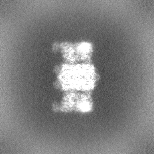
| Images | 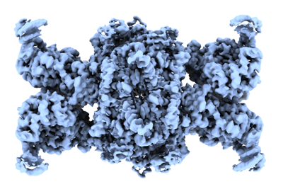 emd_19492.png emd_19492.png | 140.9 KB | ||
| Masks | emd_19492_msk_1.map | 600.7 MB | Mask map | |
| Filedesc metadata | emd-19492.cif.gz | 6.6 KB | ||
| Others | emd_19492_half_map_1.map.gzemd_19492_half_map_2.map.gz | 558 MB 558.1 MB | ||
| Archive directory |  http://ftp.pdbj.org/pub/emdb/structures/EMD-19492ftp://ftp.pdbj.org/pub/emdb/structures/EMD-19492 http://ftp.pdbj.org/pub/emdb/structures/EMD-19492ftp://ftp.pdbj.org/pub/emdb/structures/EMD-19492 | HTTPS FTP |
-Related structure data
| Related structure data |  8rthMC M: atomic model generated by this map C: citing same article ( |
|---|---|
| Similar structure data |
-Links
| EMDB pages | EMDB (EBI/PDBe) / EMDataResource |
|---|---|
| Related items in Molecule of the Month |


-Map
| File | Download / File: emd_19492.map.gz / Format: CCP4 / Size: 600.7 MB / Type: IMAGE STORED AS FLOATING POINT NUMBER (4 BYTES) | ||||||||||||||||||||||||||||||||||||
|---|---|---|---|---|---|---|---|---|---|---|---|---|---|---|---|---|---|---|---|---|---|---|---|---|---|---|---|---|---|---|---|---|---|---|---|---|---|
| Annotation | volume map sharp | ||||||||||||||||||||||||||||||||||||
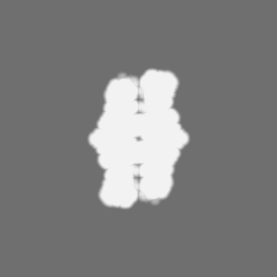
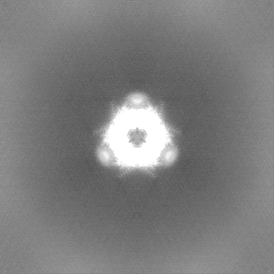
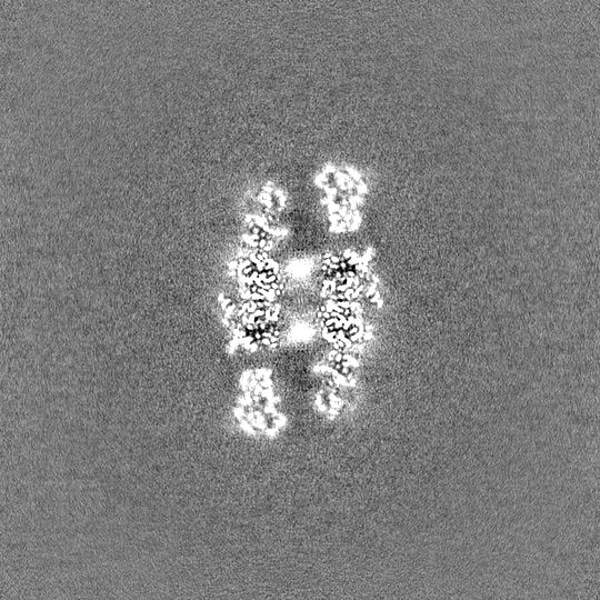
| Projections & slices | Image control
Images are generated by Spider. | ||||||||||||||||||||||||||||||||||||
| Voxel size | X=Y=Z: 0.8238 Å | ||||||||||||||||||||||||||||||||||||
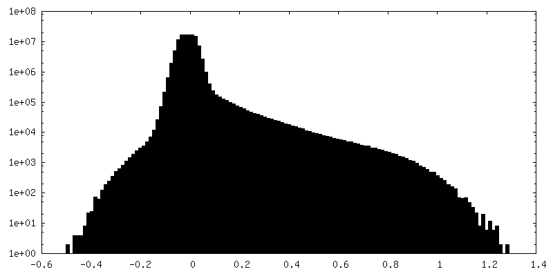
| Density |
| ||||||||||||||||||||||||||||||||||||
| Symmetry | Space group: 1 | ||||||||||||||||||||||||||||||||||||
| Details | EMDB XML:
|
 X (Sec.)
 Y (Row.)
 Z (Col.)


-Supplemental data
-Mask #1
| File | emd_19492_msk_1.map | ||||||||||||
|---|---|---|---|---|---|---|---|---|---|---|---|---|---|
| Projections & Slices |
| ||||||||||||
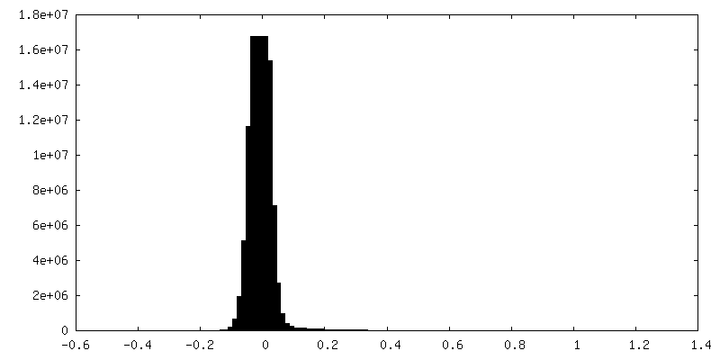
| Density Histograms |


-Half map: #2
| File | emd_19492_half_map_1.map | ||||||||||||
|---|---|---|---|---|---|---|---|---|---|---|---|---|---|
| Projections & Slices |
| ||||||||||||
| Density Histograms |


-Half map: #1
| File | emd_19492_half_map_2.map | ||||||||||||
|---|---|---|---|---|---|---|---|---|---|---|---|---|---|
| Projections & Slices |
| ||||||||||||
| Density Histograms |


- Sample components
-Entire : 3-methylcrotonyl-CoA carboxylase
| Entire | Name: 3-methylcrotonyl-CoA carboxylase |
|---|---|
| Components |
|
-Supramolecule #1: 3-methylcrotonyl-CoA carboxylase
| Supramolecule | Name: 3-methylcrotonyl-CoA carboxylase / type: complex / ID: 1 / Parent: 0 / Macromolecule list: #1-#2 |
|---|---|
| Source (natural) | Organism: Trypanosoma brucei (eukaryote) |
-Macromolecule #1: 3-methylcrotonyl-CoA carboxylase, putative
| Macromolecule | Name: 3-methylcrotonyl-CoA carboxylase, putative / type: protein_or_peptide / ID: 1 / Number of copies: 6 / Enantiomer: LEVO |
|---|---|
| Source (natural) | Organism: Trypanosoma brucei (eukaryote) |
| Molecular weight | Theoretical: 73.987648 KDa |
| Sequence | String: MLRYNVFYHG DFKKVLVANR GEIACRVFRT CREMNIRTVA VCCEGEPNAK HVLEADEAFV LGPPPASTSY LRGDRIICAA KKLQADAVH PGYGFLSENA EFASAVLAAG LKFVGPPPAA MLSMGSKSES KRIMEAAGVP IVPGYYGEDQ NPDRLLHEAK T IGFPVLIK ...String: MLRYNVFYHG DFKKVLVANR GEIACRVFRT CREMNIRTVA VCCEGEPNAK HVLEADEAFV LGPPPASTSY LRGDRIICAA KKLQADAVH PGYGFLSENA EFASAVLAAG LKFVGPPPAA MLSMGSKSES KRIMEAAGVP IVPGYYGEDQ NPDRLLHEAK T IGFPVLIK AVSGGGGKGM KIVMEETEFH LMLESAKREA INFFKDDRVI LERYVMHPRH IECQIFFDSF GNGVFFFERD CS VQRRHQK VIEEAPAPGL SVDMRRRIGD VALTAARAVG YVGAGTVEFI FDTEKDEFFF MEMNTRLQVE HPVTEQVCQV RGR PLDLVR LQLQTAMGLP LGFRQEDISM SGASVEARIY AESPRNGFLP VGGRLRYLKE PPQGNRGTVK VRLDTGFRAG DDVL VHYDP MIAKLVVWGD NRATALEGLR TALASYHIVG VETNIDFLQC CLSNPGFVEG GVTTRFIEDN SVNLLQPREI PNNVL ALAA VSYLCSQRGT STLFWPNRQI SQGVCFTVGG NPVVVRVTVS TKMCFTCDFD SSSVTVYVES TTNMPDSSTF IRVTVD GET RFGFTSFVTD SEVAVALPQG FYTLALQPLA TDFGSTSAQA NGSASVLSPM PGKVTKLLVA DGTLVQQGQA ILILEAM KM EHVVKASCDG EVKFCVHADG IVGGSTLLAH IASAAV UniProtKB: 3-methylcrotonyl-CoA carboxylase, putative |
-Macromolecule #2: methylcrotonoyl-CoA carboxylase
| Macromolecule | Name: methylcrotonoyl-CoA carboxylase / type: protein_or_peptide / ID: 2 / Number of copies: 6 / Enantiomer: LEVO / EC number: methylcrotonoyl-CoA carboxylase |
|---|---|
| Source (natural) | Organism: Trypanosoma brucei (eukaryote) |
| Molecular weight | Theoretical: 66.613969 KDa |
| Sequence | String: MKSFCRLGKV CGCSVSVVFS HRVFALGPRR DYSTSEVPLG SSQVPKGDPR KEQKGGNMSE VYLFHPAQYE SAPATTRPNV LHYPAESTN PEFKANTERM KALTAELRRR VQVIVDGDSE ADKRARDRHI SRGKLLVHQR IEKLVDPMSP FLELSQLAGG D LYPGEACH ...String: MKSFCRLGKV CGCSVSVVFS HRVFALGPRR DYSTSEVPLG SSQVPKGDPR KEQKGGNMSE VYLFHPAQYE SAPATTRPNV LHYPAESTN PEFKANTERM KALTAELRRR VQVIVDGDSE ADKRARDRHI SRGKLLVHQR IEKLVDPMSP FLELSQLAGG D LYPGEACH RGGILTGIGV VHGMRVMIVA NDATVKGGTY YPITVKKHLR AQRIAEENRL PCIYLVDSGG ANLGMQGDVF PD EQHFGRI FFNQANMSAK GIAQIATVMG SCTAGGAYVP AMSDESIIVK GNGTIFLGGP PLVFAATGEE VTPEELGGAD VHC RASGVT DYFATDDLHA LYLTRRIVAN LNRNDCERPC RGREFTPPLY DPSEIGGFIP DMGADVVKGF DVRAVIARLV DGSE FDEFK KLYGDTLVCG FARFEGMLVG IVANNGILYS ESALKGAHFV ELCSHRNIPL LFLQNITGFM VGKTYEEGGI AKNGA KLVT AVSTTHVPKI TIIIGGSYGA GNYGMCGRAF GPRFLFMWPN ARISVMGGNQ AATVLALTNS KLRENEVQDF KAKVRS KYE YEGSCYYSTA RLWDDGVIAP EDTRAVVVQA LLSTLSAPCG ETKFGVFRM UniProtKB: methylcrotonoyl-CoA carboxylase |
-Macromolecule #3: 5-(HEXAHYDRO-2-OXO-1H-THIENO[3,4-D]IMIDAZOL-6-YL)PENTANAL
| Macromolecule | Name: 5-(HEXAHYDRO-2-OXO-1H-THIENO[3,4-D]IMIDAZOL-6-YL)PENTANAL type: ligand / ID: 3 / Number of copies: 6 / Formula: BTI |
|---|---|
| Molecular weight | Theoretical: 228.311 Da |
| Chemical component information |  ChemComp-BTI: |
-Experimental details
-Structure determination
| Method | cryo EM |
|---|---|
 Processing Processing | single particle reconstruction |
| Aggregation state | particle |
-Sample preparation
| Concentration | 0.07 mg/mL | ||||||||||||||||||
|---|---|---|---|---|---|---|---|---|---|---|---|---|---|---|---|---|---|---|---|
| Buffer | pH: 7.5 Component:
| ||||||||||||||||||
| Grid | Model: C-flat-1.2/1.3 / Material: COPPER / Mesh: 300 / Support film - Material: CARBON / Support film - topology: HOLEY / Pretreatment - Type: GLOW DISCHARGE / Pretreatment - Time: 20 sec. | ||||||||||||||||||
| Vitrification | Cryogen name: ETHANE / Chamber humidity: 100 % / Chamber temperature: 283 K / Instrument: FEI VITROBOT MARK I |
- Electron microscopy
| Microscope | FEI TITAN KRIOS |
|---|---|
| Electron beam | Acceleration voltage: 300 kV / Electron source: FIELD EMISSION GUN |
| Electron optics | Illumination mode: FLOOD BEAM / Imaging mode: BRIGHT FIELDBright-field microscopy / Nominal defocus max: 3.0 µm / Nominal defocus min: 1.0 µm |
| Image recording | Film or detector model: GATAN K3 (6k x 4k) / Average electron dose: 38.3 e/Å2 |
| Experimental equipment |  Model: Titan Krios / Image courtesy: FEI Company |
-Image processing
| Particle selection | Number selected: 490000 |
|---|---|
| Startup model | Type of model: NONE |
| Initial angle assignment | Type: NOT APPLICABLE |
| Final angle assignment | Type: PROJECTION MATCHING |
| Final reconstruction | Applied symmetry - Point group: D3 (2x3 fold dihedral) / Resolution.type: BY AUTHOR / Resolution: 2.37 Å / Resolution method: FSC 0.143 CUT-OFF / Software - Name: cryoSPARC (ver. 4.4) / Number images used: 126391 |
| FSC plot (resolution estimation) | 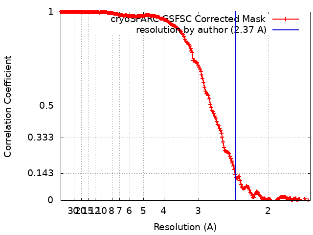 |
-Atomic model buiding 1
| Initial model | (Chain: A, AlphaFold, in silico model, B, AlphaFold, in silico model) |
|---|---|
| Refinement | Space: REAL / Protocol: FLEXIBLE FIT |
| Output model | PDB-8rth: |
