 Movie
Movie Controller

[English] 日本語
 Yorodumi
- EMDB-19425: Low-dose cryo-electron ptychographic reconstruction of apoferriti... -
+ Open data

- Basic information
| Entry |  | |||||||||
|---|---|---|---|---|---|---|---|---|---|---|
| Title | Low-dose cryo-electron ptychographic reconstruction of apoferritin recorded with CSA of 4.0 mrad | |||||||||
 Map data Map data | sharpened map | |||||||||
 Sample Sample |
| |||||||||
 Keywords Keywords |  ApoFerritin / METAL BINDING PROTEIN ApoFerritin / METAL BINDING PROTEIN | |||||||||
| Biological species |  Mus musculus (house mouse) Mus musculus (house mouse) | |||||||||
| Method | single particle reconstruction / cryo EM / Resolution: 5.8 Å | |||||||||
 Authors Authors | Mohammed I / Stalhberg H / Kucukoglu B | |||||||||
| Funding support |  Switzerland, 1 items Switzerland, 1 items
| |||||||||
 Citation Citation | Journal: To Be Published Title: Low-dose cryo-electron ptychography at sub-nanometer resolution of proteins Authors: Mohammed I / Stalhberg H / Kucukoglu B | |||||||||
| History |
|
- Structure visualization
| Supplemental images |
|---|

- Downloads & links
-EMDB archive
| Map data | emd_19425.map.gz | 13 MB |  EMDB map data format EMDB map data format | |
|---|---|---|---|---|
| Header (meta data) | emd-19425-v30.xmlemd-19425.xml | 12.3 KB 12.3 KB | Display Display | EMDB header |
| FSC (resolution estimation) | emd_19425_fsc.xml | 5.8 KB | Display | FSC data file |
| Images | 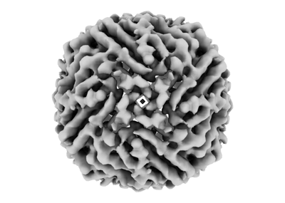 emd_19425.png emd_19425.png | 66 KB | ||
| Masks | emd_19425_msk_1.map | 13.9 MB | Mask map | |
| Filedesc metadata | emd-19425.cif.gz | 4 KB | ||
| Others | emd_19425_half_map_1.map.gzemd_19425_half_map_2.map.gz | 12.8 MB 12.8 MB | ||
| Archive directory |  http://ftp.pdbj.org/pub/emdb/structures/EMD-19425ftp://ftp.pdbj.org/pub/emdb/structures/EMD-19425 http://ftp.pdbj.org/pub/emdb/structures/EMD-19425ftp://ftp.pdbj.org/pub/emdb/structures/EMD-19425 | HTTPS FTP |
-Related structure data


-Links
| EMDB pages | EMDB (EBI/PDBe) / EMDataResource |
|---|
-Map
| File | Download / File: emd_19425.map.gz / Format: CCP4 / Size: 13.9 MB / Type: IMAGE STORED AS FLOATING POINT NUMBER (4 BYTES) | ||||||||||||||||||||
|---|---|---|---|---|---|---|---|---|---|---|---|---|---|---|---|---|---|---|---|---|---|
| Annotation | sharpened map | ||||||||||||||||||||
| Voxel size | X=Y=Z: 1.5 Å | ||||||||||||||||||||
| Density |
| ||||||||||||||||||||
| Symmetry | Space group: 1 | ||||||||||||||||||||
| Details | EMDB XML:
|
-Supplemental data
-Mask #1
| File | emd_19425_msk_1.map | ||||||||||||
|---|---|---|---|---|---|---|---|---|---|---|---|---|---|
| Projections & Slices |
| ||||||||||||
| Density Histograms |
 Z
Z Y
Y X
X


-Half map: #2
| File | emd_19425_half_map_1.map | ||||||||||||
|---|---|---|---|---|---|---|---|---|---|---|---|---|---|
| Projections & Slices |
| ||||||||||||
| Density Histograms |


-Half map: #1
| File | emd_19425_half_map_2.map | ||||||||||||
|---|---|---|---|---|---|---|---|---|---|---|---|---|---|
| Projections & Slices |
| ||||||||||||
| Density Histograms |


- Sample components
-Entire : Apoferritin
| Entire | Name: ApoferritinFerritin |
|---|---|
| Components |
|
-Supramolecule #1: Apoferritin
| Supramolecule | Name: Apoferritin / type: complex / ID: 1 / Parent: 0 |
|---|---|
| Source (natural) | Organism: Mus musculus (house mouse) |
| Molecular weight | Theoretical: 480 KDa |
-Experimental details
-Structure determination
| Method | cryo EM |
|---|---|
 Processing Processing | single particle reconstruction |
| Aggregation state | particle |
-Sample preparation
| Concentration | 5 mg/mL |
|---|---|
| Buffer | pH: 7.4 |
| Vitrification | Cryogen name: ETHANE / Instrument: FEI VITROBOT MARK IV |
- Electron microscopy
| Microscope | FEI TITAN KRIOS |
|---|---|
| Electron beam | Acceleration voltage: 300 kV / Electron source: FIELD EMISSION GUN |
| Electron optics | Illumination mode: OTHER / Imaging mode: OTHER / Nominal defocus max: 1.7 µm / Nominal defocus min: 1.4000000000000001 µm |
| Image recording | Film or detector model: DECTRIS ELA (1k x 0.5k) / Average electron dose: 32.0 e/Å2 |
| Experimental equipment |  Model: Titan Krios / Image courtesy: FEI Company |
-Image processing
| Startup model | Type of model: NONE / Details: ab-initio |
|---|---|
| Initial angle assignment | Type: MAXIMUM LIKELIHOOD / Software - Name: cryoSPARC (ver. 4.1) / Details: point group symmetry applied:O |
| Final angle assignment | Type: MAXIMUM LIKELIHOOD / Software - Name: cryoSPARC (ver. 4.1) / Details: point group symmetry applied:O |
| Final reconstruction | Applied symmetry - Point group: O (octahedral) / Algorithm: BACK PROJECTION / Resolution.type: BY AUTHOR / Resolution: 5.8 Å / Resolution method: FSC 0.143 CUT-OFF / Software - Name: cryoSPARC (ver. 4.1) / Number images used: 11552 |
| Details | py4DSTEM cryoSPARC |
| FSC plot (resolution estimation) |  |
