- EMDB-19194: In situ cryo-electron tomogram of an autophagosome in the project... -
+
Open data
ID or keywords:
Loading...
-
Basic information
Entry
Database: EMDB / ID: EMD-19194
Title
In situ cryo-electron tomogram of an autophagosome in the projection of an iPSC-derived neuron #2
Map data
Tomogram of an autophagosome containing ER captured in the projection of an iPSC-derived neuron. Tomogram denoised with cryo-CARE with a model trained on this same tomogram.
Sample
Organelle or cellular component: autophagosome containing membrane cargo captured in situ in the peripheral projections of iPSC-derived neurons grown a EM grid
Keywords
Autophagy / ERphagy / Degradation / Neuron / Cargo / Microtubules / ER / Autophagosome / CYTOSOLIC PROTEIN
Journal: Nat Cell Biol / Year: 2024 Title: Combinatorial selective ER-phagy remodels the ER during neurogenesis. Authors: Melissa J Hoyer / Cristina Capitanio / Ian R Smith / Julia C Paoli / Anna Bieber / Yizhi Jiang / Joao A Paulo / Miguel A Gonzalez-Lozano / Wolfgang Baumeister / Florian Wilfling / Brenda A ...Authors: Melissa J Hoyer / Cristina Capitanio / Ian R Smith / Julia C Paoli / Anna Bieber / Yizhi Jiang / Joao A Paulo / Miguel A Gonzalez-Lozano / Wolfgang Baumeister / Florian Wilfling / Brenda A Schulman / J Wade Harper / Abstract: The endoplasmic reticulum (ER) employs a diverse proteome landscape to orchestrate many cellular functions, ranging from protein and lipid synthesis to calcium ion flux and inter-organelle ...The endoplasmic reticulum (ER) employs a diverse proteome landscape to orchestrate many cellular functions, ranging from protein and lipid synthesis to calcium ion flux and inter-organelle communication. A case in point concerns the process of neurogenesis, where a refined tubular ER network is assembled via ER shaping proteins into the newly formed neuronal projections to create highly polarized dendrites and axons. Previous studies have suggested a role for autophagy in ER remodelling, as autophagy-deficient neurons in vivo display axonal ER accumulation within synaptic boutons, and the membrane-embedded ER-phagy receptor FAM134B has been genetically linked with human sensory and autonomic neuropathy. However, our understanding of the mechanisms underlying selective removal of the ER and the role of individual ER-phagy receptors is limited. Here we combine a genetically tractable induced neuron (iNeuron) system for monitoring ER remodelling during in vitro differentiation with proteomic and computational tools to create a quantitative landscape of ER proteome remodelling via selective autophagy. Through analysis of single and combinatorial ER-phagy receptor mutants, we delineate the extent to which each receptor contributes to both the magnitude and selectivity of ER protein clearance. We define specific subsets of ER membrane or lumenal proteins as preferred clients for distinct receptors. Using spatial sensors and flux reporters, we demonstrate receptor-specific autophagic capture of ER in axons, and directly visualize tubular ER membranes within autophagosomes in neuronal projections by cryo-electron tomography. This molecular inventory of ER proteome remodelling and versatile genetic toolkit provide a quantitative framework for understanding the contributions of individual ER-phagy receptors for reshaping ER during cell state transitions.
Download / File: emd_19194.map.gz / Format: CCP4 / Size: 2.8 GB / Type: IMAGE STORED AS FLOATING POINT NUMBER (4 BYTES)
Annotation
Tomogram of an autophagosome containing ER captured in the projection of an iPSC-derived neuron. Tomogram denoised with cryo-CARE with a model trained on this same tomogram.
Voxel size
X=Y=Z: 11.72 Å
Density
Minimum - Maximum
-20.884499999999999 - 13.985004999999999
Average (Standard dev.)
0.15032952 (±0.8427676)
Symmetry
Space group: 1
Details
EMDB XML:
Map geometry
Axis order
X
Y
Z
Origin
0
0
0
Dimensions
1024
1024
712
Spacing
1024
1024
712
Cell
A: 12001.28 Å / B: 12001.28 Å / C: 8344.641 Å α=β=γ: 90.0 °
-
Supplemental data
-
Sample components
-
Entire : autophagosome containing membrane cargo captured in situ in the p...
Entire
Name: autophagosome containing membrane cargo captured in situ in the peripheral projections of iPSC-derived neurons grown a EM grid
Components
Organelle or cellular component: autophagosome containing membrane cargo captured in situ in the peripheral projections of iPSC-derived neurons grown a EM grid
-
Supramolecule #1: autophagosome containing membrane cargo captured in situ in the p...
Supramolecule
Name: autophagosome containing membrane cargo captured in situ in the peripheral projections of iPSC-derived neurons grown a EM grid type: organelle_or_cellular_component / ID: 1 / Parent: 0
Source (natural)
Organism: Homo sapiens (human) / Strain: iPSC KOLF2.0_AAVS-TREG3-NGN2 / Tissue: induced-Neurons, DIV 18 / Location in cell: neuronal projections
-
Experimental details
-
Structure determination
Method
cryo EM
Processing
electron tomography
Aggregation state
cell
-
Sample preparation
Buffer
pH: 7
Grid
Model: Quantifoil R2/1 / Material: GOLD / Mesh: 200 / Support film - topology: HOLEY / Pretreatment - Type: PLASMA CLEANING / Pretreatment - Time: 45 sec. / Details: Grid coated with Matrigel
Vitrification
Cryogen name: ETHANE-PROPANE / Chamber humidity: 70 % / Instrument: FEI VITROBOT MARK IV
Details
iPSC-derived iNeurons on the grid were transduced at Day 12 with Lentiviruses carrying mCherry-LC3B and TEX264-GFP and plunged at DIV 18.
Cryo protectant
none
Sectioning
Other: NO SECTIONING
-
Electron microscopy
Microscope
TFS KRIOS
Electron beam
Acceleration voltage: 300 kV / Electron source: FIELD EMISSION GUN
In the structure databanks used in Yorodumi, some data are registered as the other names, "COVID-19 virus" and "2019-nCoV". Here are the details of the virus and the list of structure data.
Jan 31, 2019. EMDB accession codes are about to change! (news from PDBe EMDB page)
EMDB accession codes are about to change! (news from PDBe EMDB page)
The allocation of 4 digits for EMDB accession codes will soon come to an end. Whilst these codes will remain in use, new EMDB accession codes will include an additional digit and will expand incrementally as the available range of codes is exhausted. The current 4-digit format prefixed with “EMD-” (i.e. EMD-XXXX) will advance to a 5-digit format (i.e. EMD-XXXXX), and so on. It is currently estimated that the 4-digit codes will be depleted around Spring 2019, at which point the 5-digit format will come into force.
The EM Navigator/Yorodumi systems omit the EMD- prefix.
Related info.:Q: What is EMD? / ID/Accession-code notation in Yorodumi/EM Navigator
Yorodumi is a browser for structure data from EMDB, PDB, SASBDB, etc.
This page is also the successor to EM Navigator detail page, and also detail information page/front-end page for Omokage search.
The word "yorodu" (or yorozu) is an old Japanese word meaning "ten thousand". "mi" (miru) is to see.
Related info.:EMDB / PDB / SASBDB / Comparison of 3 databanks / Yorodumi Search / Aug 31, 2016. New EM Navigator & Yorodumi / Yorodumi Papers / Jmol/JSmol / Function and homology information / Changes in new EM Navigator and Yorodumi
 Movie
Movie Controller

 Yorodumi
 Open data
 Basic information
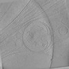
 Map data
 Sample
 Keywords
 Autophagy / ERphagy / Degradation /
 Authors
 United States, 1 items
  Citation
 Structure visualization
 Downloads & links
 EMDB map data format
 emd_19194.png
 http://ftp.pdbj.org/pub/emdb/structures/EMD-19194
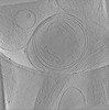
 Sample components
 Processing
 Electron microscopy
