 Movie
Movie Controller


+ Open data

- Basic information
| Entry |  | |||||||||
|---|---|---|---|---|---|---|---|---|---|---|
| Title | Cryo-EM structure of the cross-exon pre-B+ATP complex | |||||||||
 Map data Map data | ||||||||||
 Sample Sample |
| |||||||||
 Keywords Keywords |  spliceosome / SPLICING spliceosome / SPLICING | |||||||||
| Function / homology |  Function and homology informationLsm2-8 complex / U6 snRNA 3'-end binding / spliceosomal snRNP complex / ribonucleoprotein complex localization / U4atac snRNP / positive regulation of cytotoxic T cell differentiation / maturation of 5S rRNA / RNA localization / R-loop processing / positive regulation of primary miRNA processing ...Lsm2-8 complex / U6 snRNA 3'-end binding / spliceosomal snRNP complex / ribonucleoprotein complex localization / U4atac snRNP / positive regulation of cytotoxic T cell differentiation / maturation of 5S rRNA / RNA localization / R-loop processing / positive regulation of primary miRNA processing / U4atac snRNA binding / mRNA decay by 5' to 3' exoribonuclease / Lsm1-7-Pat1 complex / U11/U12 snRNP / U6 snRNP / box C/D sno(s)RNA binding / PH domain binding / snRNP binding / U2 snRNP binding / U7 snRNA binding / histone pre-mRNA DCP binding / U7 snRNP / dense fibrillar component / B-WICH complex / histone pre-mRNA 3'end processing complex / cis assembly of pre-catalytic spliceosome / SLBP independent Processing of Histone Pre-mRNAs / SLBP Dependent Processing of Replication-Dependent Histone Pre-mRNAs / splicing factor binding / spliceosome conformational change to release U4 (or U4atac) and U1 (or U11) / protein methylation / U12-type spliceosomal complex / methylosome / P-body assembly / U4/U6 snRNP / 7-methylguanosine cap hypermethylation / U1 snRNP binding / pICln-Sm protein complex / : / blastocyst formation / U2-type catalytic step 1 spliceosome / RNA splicing, via transesterification reactions / small nuclear ribonucleoprotein complex / sno(s)RNA-containing ribonucleoprotein complex / P granule / SMN-Sm protein complex / spliceosomal tri-snRNP complex / U4 snRNA binding / telomerase holoenzyme complex / box C/D methylation guide snoRNP complex / mRNA cis splicing, via spliceosome / U2-type spliceosomal complex / telomerase RNA binding / U2-type precatalytic spliceosome / commitment complex / U2-type prespliceosome assembly / U2-type catalytic step 2 spliceosome / U4 snRNP / rRNA modification in the nucleus and cytosol / box C/D snoRNP assembly / SAGA complex / positive regulation of mRNA splicing, via spliceosome / U2 snRNP / RNA Polymerase II Transcription Termination / nuclear-transcribed mRNA catabolic process / positive regulation of transcription by RNA polymerase III / U1 snRNP / U3 snoRNA binding / tRNA processing / U2-type prespliceosome / K63-linked polyubiquitin modification-dependent protein binding / cyclosporin A binding / positive regulation of miRNA metabolic process / precatalytic spliceosome / positive regulation of transcription by RNA polymerase I / spliceosomal complex assembly / regulation of alternative mRNA splicing, via spliceosome / mRNA Splicing - Minor Pathway / mRNA catabolic process / regulation of RNA splicing / mRNA 3'-splice site recognition / MLL1 complex / spliceosomal tri-snRNP complex assembly / single fertilization / Major pathway of rRNA processing in the nucleolus and cytosol / U5 snRNA binding / U5 snRNP / positive regulation of viral genome replication / RNA processing / U2 snRNA binding / Cajal body / U6 snRNA binding / regulation of DNA repair / spliceosomal snRNP assembly / protein peptidyl-prolyl isomerization / ribonucleoprotein complex binding / pre-mRNA intronic binding / U1 snRNA binding / U4/U6 x U5 tri-snRNP complex / Gene and protein expression by JAK-STAT signaling after Interleukin-12 stimulation Function and homology informationLsm2-8 complex / U6 snRNA 3'-end binding / spliceosomal snRNP complex / ribonucleoprotein complex localization / U4atac snRNP / positive regulation of cytotoxic T cell differentiation / maturation of 5S rRNA / RNA localization / R-loop processing / positive regulation of primary miRNA processing ...Lsm2-8 complex / U6 snRNA 3'-end binding / spliceosomal snRNP complex / ribonucleoprotein complex localization / U4atac snRNP / positive regulation of cytotoxic T cell differentiation / maturation of 5S rRNA / RNA localization / R-loop processing / positive regulation of primary miRNA processing / U4atac snRNA binding / mRNA decay by 5' to 3' exoribonuclease / Lsm1-7-Pat1 complex / U11/U12 snRNP / U6 snRNP / box C/D sno(s)RNA binding / PH domain binding / snRNP binding / U2 snRNP binding / U7 snRNA binding / histone pre-mRNA DCP binding / U7 snRNP / dense fibrillar component / B-WICH complex / histone pre-mRNA 3'end processing complex / cis assembly of pre-catalytic spliceosome / SLBP independent Processing of Histone Pre-mRNAs / SLBP Dependent Processing of Replication-Dependent Histone Pre-mRNAs / splicing factor binding / spliceosome conformational change to release U4 (or U4atac) and U1 (or U11) / protein methylation / U12-type spliceosomal complex / methylosome / P-body assembly / U4/U6 snRNP / 7-methylguanosine cap hypermethylation / U1 snRNP binding / pICln-Sm protein complex / : / blastocyst formation / U2-type catalytic step 1 spliceosome / RNA splicing, via transesterification reactions / small nuclear ribonucleoprotein complex / sno(s)RNA-containing ribonucleoprotein complex / P granule / SMN-Sm protein complex / spliceosomal tri-snRNP complex / U4 snRNA binding / telomerase holoenzyme complex / box C/D methylation guide snoRNP complex / mRNA cis splicing, via spliceosome / U2-type spliceosomal complex / telomerase RNA binding / U2-type precatalytic spliceosome / commitment complex / U2-type prespliceosome assembly / U2-type catalytic step 2 spliceosome / U4 snRNP / rRNA modification in the nucleus and cytosol / box C/D snoRNP assembly / SAGA complex / positive regulation of mRNA splicing, via spliceosome / U2 snRNP / RNA Polymerase II Transcription Termination / nuclear-transcribed mRNA catabolic process / positive regulation of transcription by RNA polymerase III / U1 snRNP / U3 snoRNA binding / tRNA processing / U2-type prespliceosome / K63-linked polyubiquitin modification-dependent protein binding / cyclosporin A binding / positive regulation of miRNA metabolic process / precatalytic spliceosome / positive regulation of transcription by RNA polymerase I / spliceosomal complex assembly / regulation of alternative mRNA splicing, via spliceosome / mRNA Splicing - Minor Pathway / mRNA catabolic process / regulation of RNA splicing / mRNA 3'-splice site recognition / MLL1 complex / spliceosomal tri-snRNP complex assembly / single fertilization / Major pathway of rRNA processing in the nucleolus and cytosol / U5 snRNA binding / U5 snRNP / positive regulation of viral genome replication / RNA processing / U2 snRNA binding / Cajal body / U6 snRNA binding / regulation of DNA repair / spliceosomal snRNP assembly / protein peptidyl-prolyl isomerization / ribonucleoprotein complex binding / pre-mRNA intronic binding / U1 snRNA binding / U4/U6 x U5 tri-snRNP complex / Gene and protein expression by JAK-STAT signaling after Interleukin-12 stimulationSimilarity search - Function | |||||||||
| Biological species |  Homo sapiens (human) Homo sapiens (human) | |||||||||
| Method | single particle reconstruction / cryo EM / Resolution: 4.4 Å | |||||||||
 Authors Authors | Zhang Z / Kumar V / Dybkov O / Will CL / Zhong J / Ludwig S / Urlaub H / Kastner B / Stark H / Luehrmann R | |||||||||
| Funding support |  Germany, 1 items Germany, 1 items
| |||||||||
 Citation Citation | Journal: Nature / Year: 2024 Title: Structural insights into the cross-exon to cross-intron spliceosome switch. Authors: Zhenwei Zhang / Vinay Kumar / Olexandr Dybkov / Cindy L Will / Jiayun Zhong / Sebastian E J Ludwig / Henning Urlaub / Berthold Kastner / Holger Stark / Reinhard Lührmann /  Abstract: Early spliceosome assembly can occur through an intron-defined pathway, whereby U1 and U2 small nuclear ribonucleoprotein particles (snRNPs) assemble across the intron. Alternatively, it can occur ...Early spliceosome assembly can occur through an intron-defined pathway, whereby U1 and U2 small nuclear ribonucleoprotein particles (snRNPs) assemble across the intron. Alternatively, it can occur through an exon-defined pathway, whereby U2 binds the branch site located upstream of the defined exon and U1 snRNP interacts with the 5' splice site located directly downstream of it. The U4/U6.U5 tri-snRNP subsequently binds to produce a cross-intron (CI) or cross-exon (CE) pre-B complex, which is then converted to the spliceosomal B complex. Exon definition promotes the splicing of upstream introns and plays a key part in alternative splicing regulation. However, the three-dimensional structure of exon-defined spliceosomal complexes and the molecular mechanism of the conversion from a CE-organized to a CI-organized spliceosome, a pre-requisite for splicing catalysis, remain poorly understood. Here cryo-electron microscopy analyses of human CE pre-B complex and B-like complexes reveal extensive structural similarities with their CI counterparts. The results indicate that the CE and CI spliceosome assembly pathways converge already at the pre-B stage. Add-back experiments using purified CE pre-B complexes, coupled with cryo-electron microscopy, elucidate the order of the extensive remodelling events that accompany the formation of B complexes and B-like complexes. The molecular triggers and roles of B-specific proteins in these rearrangements are also identified. We show that CE pre-B complexes can productively bind in trans to a U1 snRNP-bound 5' splice site. Together, our studies provide new mechanistic insights into the CE to CI switch during spliceosome assembly and its effect on pre-mRNA splice site pairing at this stage. | |||||||||
| History |
|
- Structure visualization
| Supplemental images |
|---|

- Downloads & links
-EMDB archive
| Map data | emd_18789.map.gz | 330.8 MB | EMDB map data format | |
|---|---|---|---|---|
| Header (meta data) | emd-18789-v30.xmlemd-18789.xml | 66.2 KB 66.2 KB | Display Display | EMDB header |
| Images | 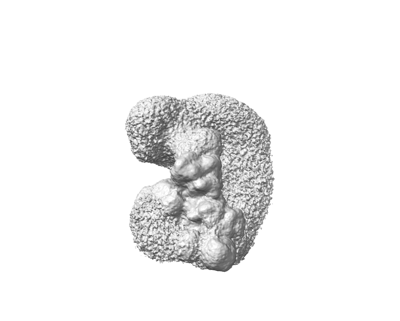 emd_18789.png emd_18789.png | 44.3 KB | ||
| Filedesc metadata | emd-18789.cif.gz | 19.8 KB | ||
| Others | emd_18789_half_map_1.map.gzemd_18789_half_map_2.map.gz | 335.6 MB 335.6 MB | ||
| Archive directory |  http://ftp.pdbj.org/pub/emdb/structures/EMD-18789ftp://ftp.pdbj.org/pub/emdb/structures/EMD-18789 http://ftp.pdbj.org/pub/emdb/structures/EMD-18789ftp://ftp.pdbj.org/pub/emdb/structures/EMD-18789 | HTTPS FTP |
-Related structure data
| Related structure data | 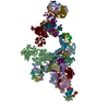 8r0bMC 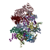 8qozC 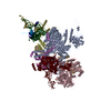 8qp8C 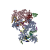 8qp9C 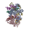 8qpaC  8qpbC 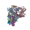 8qpeC  8qpkC 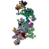 8qxdC 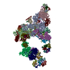 8qzsC 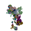 8r08C 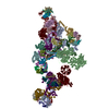 8r09C 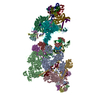 8r0aC  8rm5C M: atomic model generated by this map C: citing same article ( |
|---|---|
| Similar structure data |


-Links
| EMDB pages | EMDB (EBI/PDBe) / EMDataResource |
|---|---|
| Related items in Molecule of the Month |


-Map
| File | Download / File: emd_18789.map.gz / Format: CCP4 / Size: 421.9 MB / Type: IMAGE STORED AS FLOATING POINT NUMBER (4 BYTES) | ||||||||||||||||||||
|---|---|---|---|---|---|---|---|---|---|---|---|---|---|---|---|---|---|---|---|---|---|
| Voxel size | X=Y=Z: 1.16 Å | ||||||||||||||||||||
| Density |
| ||||||||||||||||||||
| Symmetry | Space group: 1 | ||||||||||||||||||||
| Details | EMDB XML:
|
-Supplemental data
-Half map: #2
| File | emd_18789_half_map_1.map | ||||||||||||
|---|---|---|---|---|---|---|---|---|---|---|---|---|---|
| Projections & Slices |
| ||||||||||||
| Density Histograms |
 Z
Z Y
Y X
X


-Half map: #1
| File | emd_18789_half_map_2.map | ||||||||||||
|---|---|---|---|---|---|---|---|---|---|---|---|---|---|
| Projections & Slices |
| ||||||||||||
| Density Histograms |


- Sample components
+Entire : human spliceosomal pre-B+ATP complex
+Supramolecule #1: human spliceosomal pre-B+ATP complex
+Macromolecule #1: Pre-mRNA-processing-splicing factor 8
+Macromolecule #2: U5 small nuclear ribonucleoprotein 200 kDa helicase
+Macromolecule #7: Splicing factor 3A subunit 1
+Macromolecule #8: Splicing factor 3A subunit 2
+Macromolecule #9: 116 kDa U5 small nuclear ribonucleoprotein component
+Macromolecule #10: Thioredoxin-like protein 4A
+Macromolecule #11: U5 small nuclear ribonucleoprotein 40 kDa protein
+Macromolecule #12: Probable ATP-dependent RNA helicase DDX23
+Macromolecule #13: NHP2-like protein 1, N-terminally processed
+Macromolecule #14: Ubiquitin carboxyl-terminal hydrolase 39
+Macromolecule #15: Peptidyl-prolyl cis-trans isomerase H
+Macromolecule #18: U6 snRNA-associated Sm-like protein LSm6
+Macromolecule #19: U6 snRNA-associated Sm-like protein LSm7
+Macromolecule #20: U6 snRNA-associated Sm-like protein LSm2
+Macromolecule #21: U6 snRNA-associated Sm-like protein LSm3
+Macromolecule #22: U6 snRNA-associated Sm-like protein LSm8
+Macromolecule #23: U6 snRNA-associated Sm-like protein LSm4
+Macromolecule #24: U6 snRNA-associated Sm-like protein LSm5
+Macromolecule #25: Splicing factor 3B subunit 4
+Macromolecule #26: Splicing factor 3A subunit 3
+Macromolecule #27: Splicing factor 3B subunit 2
+Macromolecule #28: Splicing factor 3B subunit 5
+Macromolecule #29: Splicing factor 3B subunit 3
+Macromolecule #30: PHD finger-like domain-containing protein 5A
+Macromolecule #31: Splicing factor 3B subunit 1
+Macromolecule #32: Splicing factor 3B subunit 6
+Macromolecule #33: Small nuclear ribonucleoprotein Sm D2
+Macromolecule #34: U2 small nuclear ribonucleoprotein B''
+Macromolecule #35: Small nuclear ribonucleoprotein F
+Macromolecule #36: Small nuclear ribonucleoprotein-associated proteins B and B'
+Macromolecule #37: Small nuclear ribonucleoprotein Sm D3
+Macromolecule #38: Small nuclear ribonucleoprotein G
+Macromolecule #39: Small nuclear ribonucleoprotein E
+Macromolecule #40: Small nuclear ribonucleoprotein Sm D1
+Macromolecule #41: U2 small nuclear ribonucleoprotein A'
+Macromolecule #42: U4/U6 small nuclear ribonucleoprotein Prp4
+Macromolecule #43: U4/U6 small nuclear ribonucleoprotein Prp3
+Macromolecule #44: U4/U6 small nuclear ribonucleoprotein Prp31
+Macromolecule #45: Pre-mRNA-processing factor 6
+Macromolecule #46: U4/U6.U5 tri-snRNP-associated protein 1
+Macromolecule #3: U2 snRNA
+Macromolecule #4: U4 snRNA
+Macromolecule #5: U5 snRNA
+Macromolecule #6: U6 snRNA
+Macromolecule #16: pre-mRNA
+Macromolecule #17: 5'ss oligo
-Experimental details
-Structure determination
| Method | cryo EM |
|---|---|
 Processing Processing | single particle reconstruction |
| Aggregation state | particle |
-Sample preparation
| Buffer | pH: 7.9 |
|---|---|
| Vitrification | Cryogen name: ETHANE |
- Electron microscopy
| Microscope | FEI TITAN KRIOS |
|---|---|
| Electron beam | Acceleration voltage: 300 kV / Electron source: FIELD EMISSION GUN |
| Electron optics | Illumination mode: FLOOD BEAM / Imaging mode: BRIGHT FIELDBright-field microscopy / Nominal defocus max: 5.0 µm / Nominal defocus min: 1.5 µm |
| Image recording | Film or detector model: FEI FALCON III (4k x 4k) / Average electron dose: 45.0 e/Å2 |
| Experimental equipment |  Model: Titan Krios / Image courtesy: FEI Company |
-Image processing
| Startup model | Type of model: OTHER / Details: ab initio |
|---|---|
| Initial angle assignment | Type: MAXIMUM LIKELIHOOD |
| Final angle assignment | Type: MAXIMUM LIKELIHOOD |
| Final reconstruction | Resolution.type: BY AUTHOR / Resolution: 4.4 Å / Resolution method: FSC 0.143 CUT-OFF / Number images used: 94460 |
