 Movie
Movie Controller

[English] 日本語
 Yorodumi
- EMDB-18590: Cryo-EM map of rotated SecM-stalled Escherichia coli 70S ribosome -
+ Open data

- Basic information
| Entry |  | |||||||||
|---|---|---|---|---|---|---|---|---|---|---|
| Title | Cryo-EM map of rotated SecM-stalled Escherichia coli 70S ribosome | |||||||||
 Map data Map data | ||||||||||
 Sample Sample |
| |||||||||
 Keywords Keywords | SecM / SRC /  Stalling / Arrest Peptide / Arrest Motive / Stalling Motive / Stalled Ribosome Complex / SecA / Secretion Monitor / Leader Peptide / TRANSLATION / RIBOSOME Stalling / Arrest Peptide / Arrest Motive / Stalling Motive / Stalled Ribosome Complex / SecA / Secretion Monitor / Leader Peptide / TRANSLATION / RIBOSOME | |||||||||
| Biological species |  Escherichia coli BW25113 (bacteria) Escherichia coli BW25113 (bacteria) | |||||||||
| Method | single particle reconstruction / cryo EM / Resolution: 2.6 Å | |||||||||
 Authors Authors | Gersteuer F / Morici M / Wilson DN | |||||||||
| Funding support |  Germany, 1 items Germany, 1 items
| |||||||||
 Citation Citation | Journal: Nat Commun / Year: 2024 Title: The SecM arrest peptide traps a pre-peptide bond formation state of the ribosome. Authors: Felix Gersteuer / Martino Morici / Sara Gabrielli / Keigo Fujiwara / Haaris A Safdari / Helge Paternoga / Lars V Bock / Shinobu Chiba / Daniel N Wilson /  Abstract: Nascent polypeptide chains can induce translational stalling to regulate gene expression. This is exemplified by the E. coli secretion monitor (SecM) arrest peptide that induces translational ...Nascent polypeptide chains can induce translational stalling to regulate gene expression. This is exemplified by the E. coli secretion monitor (SecM) arrest peptide that induces translational stalling to regulate expression of the downstream encoded SecA, an ATPase that co-operates with the SecYEG translocon to facilitate insertion of proteins into or through the cytoplasmic membrane. Here we present the structure of a ribosome stalled during translation of the full-length E. coli SecM arrest peptide at 2.0 Å resolution. The structure reveals that SecM arrests translation by stabilizing the Pro-tRNA in the A-site, but in a manner that prevents peptide bond formation with the SecM-peptidyl-tRNA in the P-site. By employing molecular dynamic simulations, we also provide insight into how a pulling force on the SecM nascent chain can relieve the SecM-mediated translation arrest. Collectively, the mechanisms determined here for SecM arrest and relief are also likely to be applicable for a variety of other arrest peptides that regulate components of the protein localization machinery identified across a wide range of bacteria lineages. | |||||||||
| History |
|
- Structure visualization
| Supplemental images |
|---|

- Downloads & links
-EMDB archive
| Map data | emd_18590.map.gz | 193.9 MB |  EMDB map data format EMDB map data format | |
|---|---|---|---|---|
| Header (meta data) | emd-18590-v30.xmlemd-18590.xml | 21.5 KB 21.5 KB | Display Display | EMDB header |
| FSC (resolution estimation) | emd_18590_fsc.xml | 14.1 KB | Display | FSC data file |
| Images | 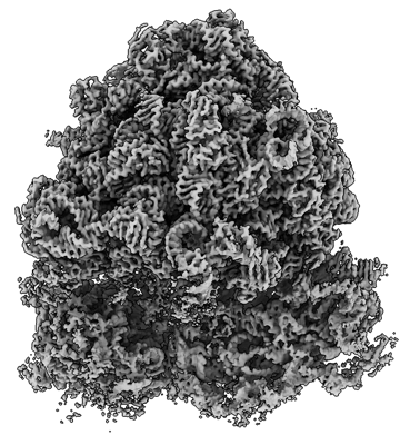 emd_18590.png emd_18590.png | 169 KB | ||
| Filedesc metadata | emd-18590.cif.gz | 4 KB | ||
| Others | emd_18590_additional_1.map.gzemd_18590_half_map_1.map.gzemd_18590_half_map_2.map.gz | 228.2 MB 194.2 MB 194.2 MB | ||
| Archive directory |  http://ftp.pdbj.org/pub/emdb/structures/EMD-18590ftp://ftp.pdbj.org/pub/emdb/structures/EMD-18590 http://ftp.pdbj.org/pub/emdb/structures/EMD-18590ftp://ftp.pdbj.org/pub/emdb/structures/EMD-18590 | HTTPS FTP |
-Related structure data

-Links
| EMDB pages | EMDB (EBI/PDBe) / EMDataResource |
|---|
-Map
| File | Download / File: emd_18590.map.gz / Format: CCP4 / Size: 244.1 MB / Type: IMAGE STORED AS FLOATING POINT NUMBER (4 BYTES) | ||||||||||||||||||||
|---|---|---|---|---|---|---|---|---|---|---|---|---|---|---|---|---|---|---|---|---|---|
| Voxel size | X=Y=Z: 0.83 Å | ||||||||||||||||||||
| Density |
| ||||||||||||||||||||
| Symmetry | Space group: 1 | ||||||||||||||||||||
| Details | EMDB XML:
|
-Supplemental data
-Additional map: #1
| File | emd_18590_additional_1.map | ||||||||||||
|---|---|---|---|---|---|---|---|---|---|---|---|---|---|
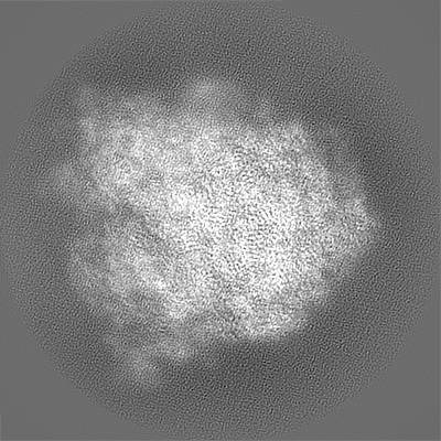
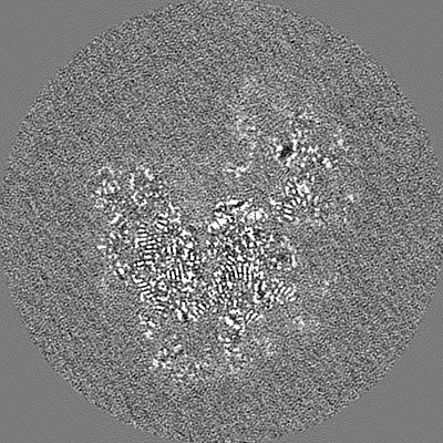
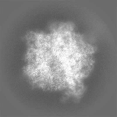
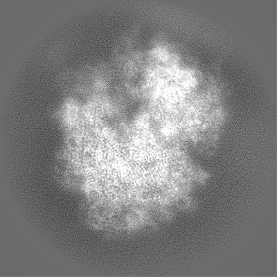
| Projections & Slices |
| ||||||||||||
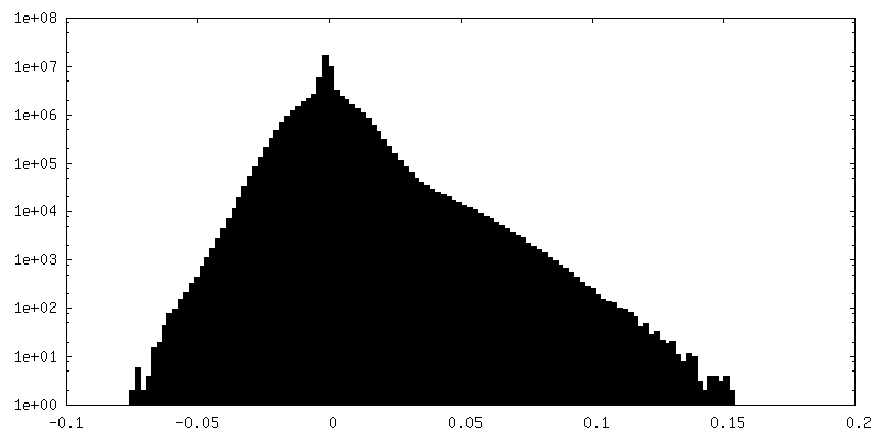
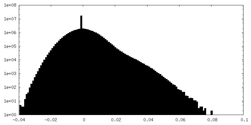
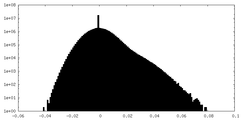
| Density Histograms |
 Z
Z Y
Y X
X


-Half map: #1
| File | emd_18590_half_map_1.map | ||||||||||||
|---|---|---|---|---|---|---|---|---|---|---|---|---|---|
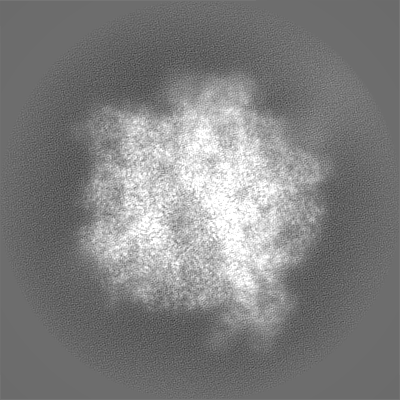
| Projections & Slices |
| ||||||||||||
| Density Histograms |


-Half map: #2
| File | emd_18590_half_map_2.map | ||||||||||||
|---|---|---|---|---|---|---|---|---|---|---|---|---|---|
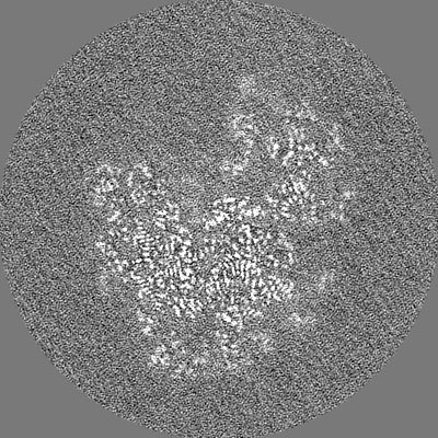
| Projections & Slices |
| ||||||||||||
| Density Histograms |


- Sample components
-Entire : SecM-stalled ribosome complex
| Entire | Name: SecM-stalled ribosome complex |
|---|---|
| Components |
|
-Supramolecule #1: SecM-stalled ribosome complex
| Supramolecule | Name: SecM-stalled ribosome complex / type: complex / ID: 1 / Parent: 0 / Macromolecule list: #1-#57 |
|---|---|
| Source (natural) | Organism: Escherichia coli BW25113 (bacteria) |
-Experimental details
-Structure determination
| Method | cryo EM |
|---|---|
 Processing Processing | single particle reconstruction |
| Aggregation state | particle |
-Sample preparation
| Buffer | pH: 7.4 |
|---|---|
| Vitrification | Cryogen name: ETHANE-PROPANE |
- Electron microscopy
| Microscope | FEI TITAN KRIOS |
|---|---|
| Electron beam | Acceleration voltage: 300 kV / Electron source: FIELD EMISSION GUN |
| Electron optics | Illumination mode: FLOOD BEAM / Imaging mode: BRIGHT FIELDBright-field microscopy / Nominal defocus max: 0.9 µm / Nominal defocus min: 0.3 µm |
| Image recording | Film or detector model: GATAN K3 BIOQUANTUM (6k x 4k) / Average electron dose: 40.0 e/Å2 |
| Experimental equipment |  Model: Titan Krios / Image courtesy: FEI Company |
-Image processing
| Startup model | Type of model: PDB ENTRY PDB model - PDB ID: |
|---|---|
| Initial angle assignment | Type: MAXIMUM LIKELIHOOD |
| Final angle assignment | Type: MAXIMUM LIKELIHOOD |
| Final reconstruction | Resolution.type: BY AUTHOR / Resolution: 2.6 Å / Resolution method: FSC 0.143 CUT-OFF / Number images used: 36489 |
| FSC plot (resolution estimation) |  |

