 Movie
Movie Controller


+ Open data

- Basic information
| Entry | 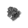 | |||||||||
|---|---|---|---|---|---|---|---|---|---|---|
| Title | ApdA-SRC with P-tRNA only | |||||||||
 Map data Map data | Postprocessed final map | |||||||||
 Sample Sample |
| |||||||||
 Keywords Keywords |  Stalling / Nascent chain / elongation arrest / regulation / RIBOSOME Stalling / Nascent chain / elongation arrest / regulation / RIBOSOME | |||||||||
| Biological species |  Amycolatopsis japonica (bacteria) Amycolatopsis japonica (bacteria) | |||||||||
| Method | single particle reconstruction / cryo EM / Resolution: 2.3 Å | |||||||||
 Authors Authors | Morici M / Wilson DN | |||||||||
| Funding support |  Germany, 1 items Germany, 1 items
| |||||||||
 Citation Citation | Journal: Nat Commun / Year: 2024 Title: RAPP-containing arrest peptides induce translational stalling by short circuiting the ribosomal peptidyltransferase activity. Authors: Martino Morici / Sara Gabrielli / Keigo Fujiwara / Helge Paternoga / Bertrand Beckert / Lars V Bock / Shinobu Chiba / Daniel N Wilson /  Abstract: Arrest peptides containing RAPP (ArgAlaProPro) motifs have been discovered in both Gram-positive and Gram-negative bacteria, where they are thought to regulate expression of important protein ...Arrest peptides containing RAPP (ArgAlaProPro) motifs have been discovered in both Gram-positive and Gram-negative bacteria, where they are thought to regulate expression of important protein localization machinery components. Here we determine cryo-EM structures of ribosomes stalled on RAPP arrest motifs in both Bacillus subtilis and Escherichia coli. Together with molecular dynamics simulations, our structures reveal that the RAPP motifs allow full accommodation of the A-site tRNA, but prevent the subsequent peptide bond from forming. Our data support a model where the RAP in the P-site interacts and stabilizes a single hydrogen atom on the Pro-tRNA in the A-site, thereby preventing an optimal geometry for the nucleophilic attack required for peptide bond formation to occur. This mechanism to short circuit the ribosomal peptidyltransferase activity is likely to operate for the majority of other RAPP-like arrest peptides found across diverse bacterial phylogenies. | |||||||||
| History |
|
- Structure visualization
| Supplemental images |
|---|

- Downloads & links
-EMDB archive
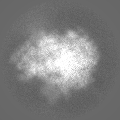
| Map data | emd_18341.map.gz | 54.7 MB |  EMDB map data format EMDB map data format | |
|---|---|---|---|---|
| Header (meta data) | emd-18341-v30.xmlemd-18341.xml | 20.9 KB 20.9 KB | Display Display | EMDB header |
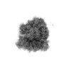
| Images | 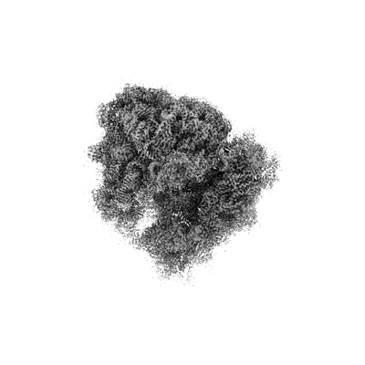 emd_18341.png emd_18341.png | 70.8 KB | ||
| Filedesc metadata | emd-18341.cif.gz | 4.1 KB | ||
| Others | emd_18341_additional_1.map.gzemd_18341_half_map_1.map.gzemd_18341_half_map_2.map.gz | 225.4 MB 225.9 MB 226 MB | ||
| Archive directory |  http://ftp.pdbj.org/pub/emdb/structures/EMD-18341ftp://ftp.pdbj.org/pub/emdb/structures/EMD-18341 http://ftp.pdbj.org/pub/emdb/structures/EMD-18341ftp://ftp.pdbj.org/pub/emdb/structures/EMD-18341 | HTTPS FTP |
-Related structure data


-Links
| EMDB pages | EMDB (EBI/PDBe) / EMDataResource |
|---|
-Map
| File | Download / File: emd_18341.map.gz / Format: CCP4 / Size: 282.6 MB / Type: IMAGE STORED AS FLOATING POINT NUMBER (4 BYTES) | ||||||||||||||||||||
|---|---|---|---|---|---|---|---|---|---|---|---|---|---|---|---|---|---|---|---|---|---|
| Annotation | Postprocessed final map | ||||||||||||||||||||
| Voxel size | X=Y=Z: 0.82 Å | ||||||||||||||||||||
| Density |
| ||||||||||||||||||||
| Symmetry | Space group: 1 | ||||||||||||||||||||
| Details | EMDB XML:
|
-Supplemental data
-Additional map: Preprocessed map
| File | emd_18341_additional_1.map | ||||||||||||
|---|---|---|---|---|---|---|---|---|---|---|---|---|---|
| Annotation | Preprocessed map | ||||||||||||
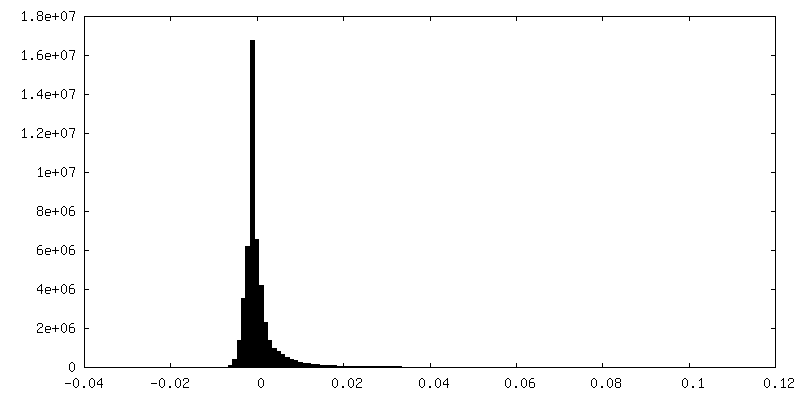
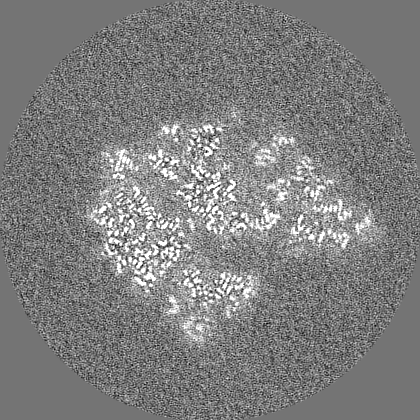
| Projections & Slices |
| ||||||||||||
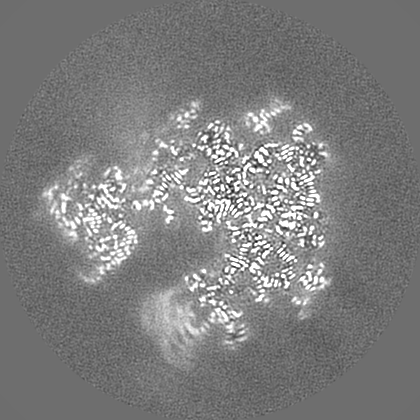
| Density Histograms |
 Z
Z Y
Y X
X


-Half map: Half-map 1
| File | emd_18341_half_map_1.map | ||||||||||||
|---|---|---|---|---|---|---|---|---|---|---|---|---|---|
| Annotation | Half-map 1 | ||||||||||||
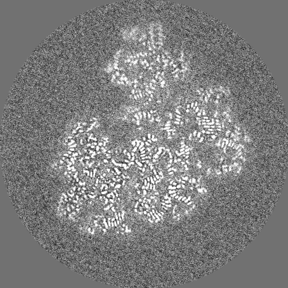
| Projections & Slices |
| ||||||||||||
| Density Histograms |


-Half map: Half-map 2
| File | emd_18341_half_map_2.map | ||||||||||||
|---|---|---|---|---|---|---|---|---|---|---|---|---|---|
| Annotation | Half-map 2 | ||||||||||||
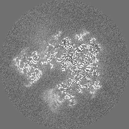
| Projections & Slices |
| ||||||||||||
| Density Histograms |


- Sample components
-Entire : B. subtilis ApdA-stalled ribosomal complex
| Entire | Name: B. subtilis ApdA-stalled ribosomal complex |
|---|---|
| Components |
|
-Supramolecule #1: B. subtilis ApdA-stalled ribosomal complex
| Supramolecule | Name: B. subtilis ApdA-stalled ribosomal complex / type: complex / ID: 1 / Parent: 0 / Macromolecule list: #1-#49 |
|---|---|
| Source (natural) | Organism: Amycolatopsis japonica (bacteria) |
-Experimental details
-Structure determination
| Method | cryo EM |
|---|---|
 Processing Processing | single particle reconstruction |
| Aggregation state | particle |
-Sample preparation
| Buffer | pH: 7.4 |
|---|---|
| Vitrification | Cryogen name: ETHANE-PROPANE |
- Electron microscopy
| Microscope | TFS KRIOS |
|---|---|
| Electron beam | Acceleration voltage: 300 kV / Electron source: FIELD EMISSION GUN |
| Electron optics | Illumination mode: FLOOD BEAM / Imaging mode: BRIGHT FIELDBright-field microscopy / Nominal defocus max: 1.8 µm / Nominal defocus min: 0.6 µm |
| Image recording | Film or detector model: GATAN K3 BIOQUANTUM (6k x 4k) / Average electron dose: 75.6 e/Å2 |
| Experimental equipment |  Model: Titan Krios / Image courtesy: FEI Company |
-Image processing
| Startup model | Type of model: INSILICO MODEL In silico model: An unpublished map from our group was initially low-pass filtered and used as model, since the biology was analogous |
|---|---|
| Initial angle assignment | Type: MAXIMUM LIKELIHOOD |
| Final angle assignment | Type: MAXIMUM LIKELIHOOD |
| Final reconstruction | Resolution.type: BY AUTHOR / Resolution: 2.3 Å / Resolution method: FSC 0.143 CUT-OFF / Number images used: 152257 |
