Movie
Movie Controller


+ Open data

- Basic information
| Entry | Database: EMDB / ID: EMD-1823 | |||||||||
|---|---|---|---|---|---|---|---|---|---|---|
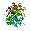
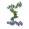
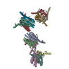
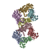
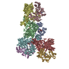
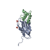
| Title | Clostridium thermocellum Mini-Cellulosome | |||||||||
 Map data Map data | This is an image of a surface rendered side-view of Clostridium thermocellum Mini-Cellulosome | |||||||||
 Sample Sample |
| |||||||||
 Keywords Keywords |  Cellulosome / CipA / Cohesin / Dockerin Cellulosome / CipA / Cohesin / Dockerin | |||||||||
| Biological species |  Clostridium thermocellum (bacteria) Clostridium thermocellum (bacteria) | |||||||||
| Method | single particle reconstruction / cryo EM / Resolution: 35.0 Å | |||||||||
 Authors Authors | Garcia-Alvarez B / Melero R / Dias FMV / Prates JAM / Fontes CMGA / Smith SP / Joao Romao M / Carvalho AL / Llorca O | |||||||||
 Citation Citation | Journal: J Mol Biol / Year: 2011 Title: Molecular architecture and structural transitions of a Clostridium thermocellum mini-cellulosome. Authors: Begoña García-Alvarez / Roberto Melero / Fernando M V Dias / José A M Prates / Carlos M G A Fontes / Steven P Smith / Maria João Romão / Ana Luísa Carvalho / Oscar Llorca /  Abstract: The cellulosome is a highly elaborate cell-bound multienzyme complex that efficiently orchestrates the deconstruction of cellulose and hemicellulose, two of the nature's most abundant polymers. ...The cellulosome is a highly elaborate cell-bound multienzyme complex that efficiently orchestrates the deconstruction of cellulose and hemicellulose, two of the nature's most abundant polymers. Understanding the intricacy of these nanomachines evolved by anaerobic microbes could sustain the development of an effective process for the conversion of lignocellulosic biomass to bio-ethanol. In Clostridium thermocellum, cellulosome assembly is mediated by high-affinity protein:protein interactions (>10(9) M(-1)) between dockerin modules found in the catalytic subunits and cohesin modules located in a non-catalytic protein scaffold termed CipA. Whereas the atomic structures of several cellulosomal components have been elucidated, the structural organization of the complete cellulosome remains elusive. Here, we reveal that a large fragment of the cellulosome presents a mostly compact conformation in solution, by solving the three-dimensional structure of a C. thermocellum mini-cellulosome comprising three consecutive cohesin modules, each bound to one Cel8A cellulase, at 35 Å resolution by cryo-electron microscopy. Interestingly, the three cellulosomal catalytic domains are found alternately projected outward from the CipA scaffold in opposite directions, in an arrangement that could expand the area of the substrate accessible to the catalytic domains. In addition, the cellulosome can transit from this compact conformation to a multitude of diverse and flexible structures, where the linkers between cohesin modules are extended and flexible. Thus, structural transitions controlled by changes in the degree of flexibility of linkers connecting consecutive cohesin modules could regulate the efficiency of substrate recognition and hydrolysis. | |||||||||
| History |
|
- Structure visualization
| Movie |
 Movie viewer Movie viewer |
|---|---|
| Structure viewer | EM map: SurfViewMolmilJmol/JSmol |
| Supplemental images |

- Downloads & links
-EMDB archive
| Map data | emd_1823.map.gz | 2.9 MB | EMDB map data format | |
|---|---|---|---|---|
| Header (meta data) | emd-1823-v30.xmlemd-1823.xml | 10.8 KB 10.8 KB | Display Display | EMDB header |
| Images |  emd_1823.jpg emd_1823.jpg | 32.4 KB | ||
| Archive directory |  http://ftp.pdbj.org/pub/emdb/structures/EMD-1823ftp://ftp.pdbj.org/pub/emdb/structures/EMD-1823 http://ftp.pdbj.org/pub/emdb/structures/EMD-1823ftp://ftp.pdbj.org/pub/emdb/structures/EMD-1823 | HTTPS FTP |
-Related structure data
| Similar structure data |
|---|

-Links
| EMDB pages | EMDB (EBI/PDBe) / EMDataResource |
|---|
-Map
| File | Download / File: emd_1823.map.gz / Format: CCP4 / Size: 4.2 MB / Type: IMAGE STORED AS FLOATING POINT NUMBER (4 BYTES) | ||||||||||||||||||||||||||||||||||||||||||||||||||||||||||||||||||||
|---|---|---|---|---|---|---|---|---|---|---|---|---|---|---|---|---|---|---|---|---|---|---|---|---|---|---|---|---|---|---|---|---|---|---|---|---|---|---|---|---|---|---|---|---|---|---|---|---|---|---|---|---|---|---|---|---|---|---|---|---|---|---|---|---|---|---|---|---|---|
| Annotation | This is an image of a surface rendered side-view of Clostridium thermocellum Mini-Cellulosome | ||||||||||||||||||||||||||||||||||||||||||||||||||||||||||||||||||||
| Voxel size | X=Y=Z: 4.2 Å | ||||||||||||||||||||||||||||||||||||||||||||||||||||||||||||||||||||
| Density |
| ||||||||||||||||||||||||||||||||||||||||||||||||||||||||||||||||||||
| Symmetry | Space group: 1 | ||||||||||||||||||||||||||||||||||||||||||||||||||||||||||||||||||||
| Details | EMDB XML:
CCP4 map header:
| ||||||||||||||||||||||||||||||||||||||||||||||||||||||||||||||||||||
-Supplemental data
- Sample components
-Entire : Mini-Cellulosome of Clostridium thermocillium
| Entire | Name: Mini-Cellulosome of Clostridium thermocillium |
|---|---|
| Components |
|
-Supramolecule #1000: Mini-Cellulosome of Clostridium thermocillium
| Supramolecule | Name: Mini-Cellulosome of Clostridium thermocillium / type: sample / ID: 1000 / Number unique components: 2 |
|---|---|
| Molecular weight | Experimental: 200 KDa |
-Macromolecule #1: Cohesin domains
| Macromolecule | Name: Cohesin domains / type: protein_or_peptide / ID: 1 / Name.synonym: C3-C4-C5 cohesin domains of CipA / Number of copies: 1 / Oligomeric state: Monomer / Recombinant expression: Yes |
|---|---|
| Source (natural) | Organism: Clostridium thermocellum (bacteria) |
| Molecular weight | Theoretical: 52.1 KDa |
| Recombinant expression | Organism: Escherichia coli (E. coli) / Recombinant plasmid: pET3a (Novagen) |
-Macromolecule #2: Clostridium thermocellum Cel8A
| Macromolecule | Name: Clostridium thermocellum Cel8A / type: protein_or_peptide / ID: 2 Name.synonym: N-terminal glycoside hydrolase family 8 (GH8) catalytic domain Number of copies: 3 / Oligomeric state: Monomer / Recombinant expression: Yes |
|---|---|
| Source (natural) | Organism: Clostridium thermocellum (bacteria) |
| Molecular weight | Theoretical: 49.9 KDa |
| Recombinant expression | Organism: Escherichia coli (E. coli) / Recombinant plasmid: pET3a (Novagen) |
-Experimental details
-Structure determination
| Method | cryo EM |
|---|---|
 Processing Processing | single particle reconstruction |
| Aggregation state | particle |
-Sample preparation
| Buffer | pH: 7.5 Details: 50 mM Hepes/HCl buffer, pH 7.5, containing 200 mM NaCl and 2 mM CaCl2 |
|---|---|
| Grid | Details: QUANTIFOIL R 1.2/1.3 |
| Vitrification | Cryogen name: ETHANE / Instrument: OTHER / Details: Vitrification instrument: FEI Vitrobot |
- Electron microscopy
| Microscope | JEOL 2200FS |
|---|---|
| Electron beam | Acceleration voltage: 200 kV / Electron source: FIELD EMISSION GUN |
| Electron optics | Calibrated magnification: 69500 / Illumination mode: FLOOD BEAM / Imaging mode: BRIGHT FIELDBright-field microscopy / Cs: 2.0 mm / Nominal magnification: 50000 |
| Specialist optics | Energy filter - Name: FEI |
| Sample stage | Specimen holder: Eucentric / Specimen holder model: GATAN LIQUID NITROGEN |
| Date | Jun 25, 2010 |
| Image recording | Category: CCD / Film or detector model: GATAN ULTRASCAN 4000 (4k x 4k) / Digitization - Sampling interval: 15 µm / Details: 0.42 nm per pixel, final sampling / Bits/pixel: 16 |
-Image processing
| CTF correction | Details: Each CCD Frame |
|---|---|
| Final reconstruction | Applied symmetry - Point group: C1 (asymmetric) / Algorithm: OTHER / Resolution.type: BY AUTHOR / Resolution: 35.0 Å / Resolution method: FSC 0.5 CUT-OFF / Software - Name: EMAN / Number images used: 5612 |
-Atomic model buiding 1
| Initial model | PDB ID: |
|---|---|
| Software | Name: Chimera |
| Details | Protocol: Rigid Body |
| Refinement | Space: REAL / Protocol: RIGID BODY FIT / Target criteria: R-factor |
-Atomic model buiding 2
| Initial model | PDB ID: |
|---|---|
| Software | Name: Chimera |
| Details | Protocol: Rigid Body |
| Refinement | Space: REAL / Protocol: RIGID BODY FIT / Target criteria: R-factor |