 Movie
Movie Controller


+ Open data

- Basic information
| Entry |  | |||||||||
|---|---|---|---|---|---|---|---|---|---|---|
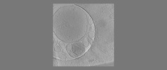
| Title | The fibrillar and amorphous states of polyQ Q97 | |||||||||
 Map data Map data | The fibrillar and amorphous states of polyQ Q97 | |||||||||
 Sample Sample |
| |||||||||
 Keywords Keywords | polyQ aggregates /  neurodegeneration / PROTEIN FIBRIL neurodegeneration / PROTEIN FIBRIL | |||||||||
| Biological species |  Homo sapiens (human) Homo sapiens (human) | |||||||||
| Method | electron tomography / cryo EM | |||||||||
 Authors Authors | Zhao DY | |||||||||
| Funding support |  United States, 1 items United States, 1 items
| |||||||||
 Citation Citation | Journal: To Be Published Title: Autophagy preferentially degrades non-fibrillar polyQ aggregates Authors: Zhao DY / Bauerlein FJB / Saha I / Hartl FU / Baumeister W / Wilfling F | |||||||||
| History |
|
- Structure visualization
| Supplemental images |
|---|

- Downloads & links
-EMDB archive
| Map data | emd_18110.map.gz | 1.4 GB |  EMDB map data format EMDB map data format | |
|---|---|---|---|---|
| Header (meta data) | emd-18110-v30.xmlemd-18110.xml | 8.3 KB 8.3 KB | Display Display | EMDB header |
| Images | 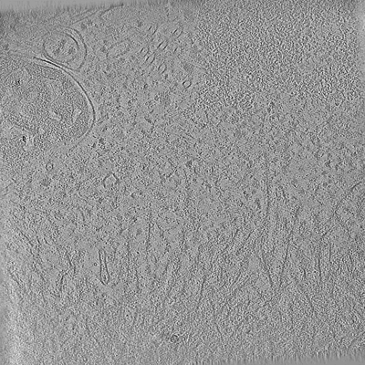 emd_18110.png emd_18110.png | 255 KB | ||
| Filedesc metadata | emd-18110.cif.gz | 3.5 KB | ||
| Archive directory |  http://ftp.pdbj.org/pub/emdb/structures/EMD-18110ftp://ftp.pdbj.org/pub/emdb/structures/EMD-18110 http://ftp.pdbj.org/pub/emdb/structures/EMD-18110ftp://ftp.pdbj.org/pub/emdb/structures/EMD-18110 | HTTPS FTP |
-Related structure data

-Links
| EMDB pages | EMDB (EBI/PDBe) / EMDataResource |
|---|
-Map
| File | Download / File: emd_18110.map.gz / Format: CCP4 / Size: 1.5 GB / Type: IMAGE STORED AS FLOATING POINT NUMBER (4 BYTES) | ||||||||||||||||||||
|---|---|---|---|---|---|---|---|---|---|---|---|---|---|---|---|---|---|---|---|---|---|
| Annotation | The fibrillar and amorphous states of polyQ Q97 | ||||||||||||||||||||
| Voxel size | X=Y=Z: 12.76 Å | ||||||||||||||||||||
| Density |
| ||||||||||||||||||||
| Symmetry | Space group: 1 | ||||||||||||||||||||
| Details | EMDB XML:
|
-Supplemental data
- Sample components
-Entire : polyQ aggregates in Hek293 cells
| Entire | Name: polyQ aggregates in Hek293 cells |
|---|---|
| Components |
|
-Supramolecule #1: polyQ aggregates in Hek293 cells
| Supramolecule | Name: polyQ aggregates in Hek293 cells / type: cell / ID: 1 / Parent: 0 |
|---|---|
| Source (natural) | Organism: Homo sapiens (human) |
-Experimental details
-Structure determination
| Method | cryo EM |
|---|---|
 Processing Processing | electron tomography |
| Aggregation state | cell |
-Sample preparation
| Buffer | pH: 7.5 / Details: Cell media with 10% glycerol |
|---|---|
| Grid | Material: GOLD / Mesh: 200 / Support film - Material: CARBON / Support film - topology: HOLEY |
| Vitrification | Cryogen name: ETHANE-PROPANE / Chamber humidity: 25 % / Chamber temperature: 298 K / Instrument: FEI VITROBOT MARK IV |
| Cryo protectant | 10% glycerol |
| Sectioning | Focused ion beam - Instrument: OTHER / Focused ion beam - Ion: OTHER / Focused ion beam - Voltage: 30 / Focused ion beam - Current: 0.05 / Focused ion beam - Duration: 1800 / Focused ion beam - Temperature: 100 K / Focused ion beam - Initial thickness: 1000 / Focused ion beam - Final thickness: 250 Focused ion beam - Details: The value given for _em_focused_ion_beam.instrument is FEI. This is not in a list of allowed values {'DB235', 'OTHER'} so OTHER is written into the XML file. |
- Electron microscopy
| Microscope | FEI TITAN KRIOS |
|---|---|
| Electron beam | Acceleration voltage: 300 kV / Electron source: FIELD EMISSION GUN |
| Electron optics | Illumination mode: FLOOD BEAM / Imaging mode: BRIGHT FIELDBright-field microscopy / Nominal defocus max: 5.0 µm / Nominal defocus min: 3.0 µm |
| Sample stage | Specimen holder model: FEI TITAN KRIOS AUTOGRID HOLDER / Cooling holder cryogen: NITROGEN |
| Image recording | Film or detector model: GATAN K2 SUMMIT (4k x 4k) / Detector mode: COUNTING / Average electron dose: 2.0 e/Å2 |
| Experimental equipment |  Model: Titan Krios / Image courtesy: FEI Company |
-Image processing
| Final reconstruction | Algorithm: BACK PROJECTION / Number images used: 50 |
|---|
