 Movie
Movie Controller

[English] 日本語
 Yorodumi
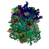
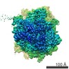
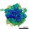
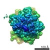
- EMDB-1811: Yeast 80S ribosome stalled by a stem-loop containing mRNA in comp... -
+ Open data

- Basic information
| Entry | Database: EMDB / ID: EMD-1811 | |||||||||
|---|---|---|---|---|---|---|---|---|---|---|
| Title | Yeast 80S ribosome stalled by a stem-loop containing mRNA in complex with Dom34-Hbs1. The dataset is computationally sorted for presence of P-site tRNA and Dom34-Hbs1. | |||||||||
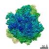
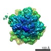
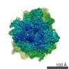
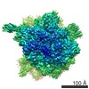
 Map data Map data | This map represents a cryo-EM reconstruction of yeast 80S ribosome stalled by a stable stem-loop structure in complex with Dom34 and Hbs1. Additionally, it contains a P-site tRNA. | |||||||||
 Sample Sample |
| |||||||||
 Keywords Keywords |  Ribosome / stalling / mRNA / P-site tRNA / no-go mRNA decay Ribosome / stalling / mRNA / P-site tRNA / no-go mRNA decay | |||||||||
| Function / homology |  Function and homology information Function and homology informationDom34-Hbs1 complex / RNA surveillance / nuclear-transcribed mRNA catabolic process, no-go decay / mRNA decay by 3' to 5' exoribonuclease / nuclear-transcribed mRNA catabolic process, non-stop decay / nonfunctional rRNA decay / ribosome disassembly / positive regulation of translational initiation / rescue of stalled ribosome / translation elongation factor activity ...Dom34-Hbs1 complex / RNA surveillance / nuclear-transcribed mRNA catabolic process, no-go decay / mRNA decay by 3' to 5' exoribonuclease / nuclear-transcribed mRNA catabolic process, non-stop decay / nonfunctional rRNA decay / ribosome disassembly / positive regulation of translational initiation / rescue of stalled ribosome / translation elongation factor activity / RNA endonuclease activity / positive regulation of translation / meiotic cell cycle / Hydrolases; Acting on acid anhydrides; Acting on GTP to facilitate cellular and subcellular movement / translation / cell division / GTPase activity / GTP binding / metal ion binding / cytosol / cytoplasmSimilarity search - Function | |||||||||
| Biological species |  Saccharomyces cerevisiae (brewer's yeast) Saccharomyces cerevisiae (brewer's yeast) | |||||||||
| Method | single particle reconstruction / cryo EM / Resolution: 9.5 Å | |||||||||
 Authors Authors | Becker T / Armache JP / Anger AM / Jarasch A / Villa E / Sieber H / AbdelMotaal B / Berninghausen O / Mielke T / Beckmann R | |||||||||
 Citation Citation | Journal: Nat Struct Mol Biol / Year: 2011 Title: Structure of the no-go mRNA decay complex Dom34-Hbs1 bound to a stalled 80S ribosome. Authors: Thomas Becker / Jean-Paul Armache / Alexander Jarasch / Andreas M Anger / Elizabeth Villa / Heidemarie Sieber / Basma Abdel Motaal / Thorsten Mielke / Otto Berninghausen / Roland Beckmann /  Abstract: No-go decay (NGD) is a mRNA quality-control mechanism in eukaryotic cells that leads to degradation of mRNAs stalled during translational elongation. The key factors triggering NGD are Dom34 and Hbs1. ...No-go decay (NGD) is a mRNA quality-control mechanism in eukaryotic cells that leads to degradation of mRNAs stalled during translational elongation. The key factors triggering NGD are Dom34 and Hbs1. We used cryo-EM to visualize NGD intermediates resulting from binding of the Dom34-Hbs1 complex to stalled ribosomes. At subnanometer resolution, all domains of Dom34 and Hbs1 were identified, allowing the docking of crystal structures and homology models. Moreover, the close structural similarity of Dom34 and Hbs1 to eukaryotic release factors (eRFs) enabled us to propose a model for the ribosome-bound eRF1-eRF3 complex. Collectively, our data provide structural insights into how stalled mRNA is recognized on the ribosome and how the eRF complex can simultaneously recognize stop codons and catalyze peptide release. | |||||||||
| History |
|
- Structure visualization
| Movie |
Movie viewer |
|---|---|
| Structure viewer | EM map: SurfViewMolmilJmol/JSmol |
| Supplemental images |

- Downloads & links
-EMDB archive
| Map data | emd_1811.map.gz | 28.9 MB | EMDB map data format | |
|---|---|---|---|---|
| Header (meta data) | emd-1811-v30.xmlemd-1811.xml | 13.8 KB 13.8 KB | Display Display | EMDB header |
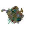
| Images | 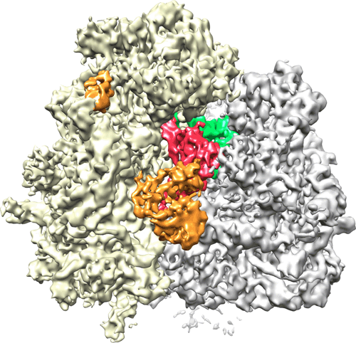 EMD-1811.gif EMD-1811.gif | 122.6 KB | ||
| Archive directory |  http://ftp.pdbj.org/pub/emdb/structures/EMD-1811ftp://ftp.pdbj.org/pub/emdb/structures/EMD-1811 http://ftp.pdbj.org/pub/emdb/structures/EMD-1811ftp://ftp.pdbj.org/pub/emdb/structures/EMD-1811 | HTTPS FTP |
-Related structure data
| Related structure data | 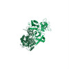 3izqMC 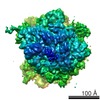 1808C  1809C 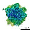 1812C M: atomic model generated by this map C: citing same article ( |
|---|---|
| Similar structure data |

-Links
| EMDB pages | EMDB (EBI/PDBe) / EMDataResource |
|---|---|
| Related items in Molecule of the Month |


-Map
| File | Download / File: emd_1811.map.gz / Format: CCP4 / Size: 185.7 MB / Type: IMAGE STORED AS FLOATING POINT NUMBER (4 BYTES) | ||||||||||||||||||||||||||||||||||||||||||||||||||||||||||||||||||||
|---|---|---|---|---|---|---|---|---|---|---|---|---|---|---|---|---|---|---|---|---|---|---|---|---|---|---|---|---|---|---|---|---|---|---|---|---|---|---|---|---|---|---|---|---|---|---|---|---|---|---|---|---|---|---|---|---|---|---|---|---|---|---|---|---|---|---|---|---|---|
| Annotation | This map represents a cryo-EM reconstruction of yeast 80S ribosome stalled by a stable stem-loop structure in complex with Dom34 and Hbs1. Additionally, it contains a P-site tRNA. | ||||||||||||||||||||||||||||||||||||||||||||||||||||||||||||||||||||
| Voxel size | X=Y=Z: 1.2375 Å | ||||||||||||||||||||||||||||||||||||||||||||||||||||||||||||||||||||
| Density |
| ||||||||||||||||||||||||||||||||||||||||||||||||||||||||||||||||||||
| Symmetry | Space group: 1 | ||||||||||||||||||||||||||||||||||||||||||||||||||||||||||||||||||||
| Details | EMDB XML:
CCP4 map header:
| ||||||||||||||||||||||||||||||||||||||||||||||||||||||||||||||||||||
-Supplemental data
- Sample components
-Entire : Stem-loop stalled yeast 80S ribosome in complex with Dom34-Hbs1 a...
| Entire | Name: Stem-loop stalled yeast 80S ribosome in complex with Dom34-Hbs1 and P-site tRNA. |
|---|---|
| Components |
|
-Supramolecule #1000: Stem-loop stalled yeast 80S ribosome in complex with Dom34-Hbs1 a...
| Supramolecule | Name: Stem-loop stalled yeast 80S ribosome in complex with Dom34-Hbs1 and P-site tRNA. type: sample / ID: 1000 Details: Mammalian Sec61 was added to saturate the hydrophobic signal sequence present in the nascent polypeptide chain. Oligomeric state: One ribosome / Number unique components: 3 |
|---|---|
| Molecular weight | Theoretical: 3.3 MDa |
-Supramolecule #1: Saccharomyces cerevisiae 80S ribosome
| Supramolecule | Name: Saccharomyces cerevisiae 80S ribosome / type: complex / ID: 1 / Name.synonym: yeast 80S ribosome Details: The mRNA stem-loop structure is not visible in the Cryo-EM reconstruction indicating its flexibility Ribosome-details: ribosome-eukaryote: ALL |
|---|---|
| Molecular weight | Experimental: 3.2 MDa / Theoretical: 3.2 MDa |
-Macromolecule #1: Hbs1p
| Macromolecule | Name: Hbs1p / type: protein_or_peptide / ID: 1 / Name.synonym: Hbs1p / Number of copies: 1 / Oligomeric state: Monomer / Recombinant expression: Yes |
|---|---|
| Source (natural) | Organism: Saccharomyces cerevisiae (brewer's yeast) / synonym: Bakers's yeast / Location in cell: cytosol |
| Molecular weight | Experimental: 68 KDa / Theoretical: 68 KDa |
| Recombinant expression | Organism:  Escherichia coli (E. coli) / Recombinant plasmid: pET28b Escherichia coli (E. coli) / Recombinant plasmid: pET28b |
-Macromolecule #2: Dom34p
| Macromolecule | Name: Dom34p / type: protein_or_peptide / ID: 2 / Name.synonym: Dom34p / Number of copies: 1 / Oligomeric state: Monomer / Recombinant expression: Yes |
|---|---|
| Source (natural) | Organism: Saccharomyces cerevisiae (brewer's yeast) / synonym: Bakers's yeast / Location in cell: cytosol |
| Molecular weight | Experimental: 44 KDa / Theoretical: 44 KDa |
| Recombinant expression | Organism: Escherichia coli (E. coli) / Recombinant plasmid: pET21a |
-Experimental details
-Structure determination
| Method | cryo EM |
|---|---|
 Processing Processing | single particle reconstruction |
| Aggregation state | particle |
-Sample preparation
| Concentration | 0.02 mg/mL |
|---|---|
| Buffer | pH: 7 Details: 20 mM Tris/HCl, pH 7.0, 80 mM NaCl, 97 mM KOAc, 10 mM Mg(OAc)2, 1.5 mM DTT, 0.02 % Nikkol, 1.8 % Glycerol, 0.01 mg/ml Cycloheximide, 500 0.5 mM GDPNP, 0.3 % Digitonin |
| Grid | Details: Quantifoil Grid with 2 nm carbon on top |
| Vitrification | Cryogen name: ETHANE / Chamber humidity: 100 % / Instrument: OTHER / Details: Vitrification instrument: Vitrobot Method: Blotted for 10 seconds before plunging, used 2 layer of filter paper |
- Electron microscopy
| Microscope | FEI POLARA 300 |
|---|---|
| Electron beam | Acceleration voltage: 300 kV / Electron source: FIELD EMISSION GUN |
| Electron optics | Calibrated magnification: 38000 / Illumination mode: FLOOD BEAM / Imaging mode: BRIGHT FIELDBright-field microscopy / Cs: 2.26 mm / Nominal defocus max: 3.78 µm / Nominal defocus min: 1.5 µm / Nominal magnification: 39000 |
| Sample stage | Specimen holder: FEI Polara Cartridge System / Specimen holder model: OTHER |
| Temperature | Average: 84 K |
| Alignment procedure | Legacy - Astigmatism: Objective lens astigmatism was corrected at 100000 times magnification |
| Image recording | Category: CCD / Film or detector model: KODAK SO-163 FILM / Digitization - Sampling interval: 4.76 µm / Number real images: 78 / Average electron dose: 25 e/Å2 Details: Scanned with a Heidelberg PrimeScan drum scanner at 5334 dpi Od range: 1.2 / Bits/pixel: 16 |
| Experimental equipment |  Model: Tecnai Polara / Image courtesy: FEI Company |
-Image processing
| CTF correction | Details: CTF correction on the level of 3D volumes (SPIDER TF CTS command) |
|---|---|
| Final reconstruction | Applied symmetry - Point group: C1 (asymmetric) / Algorithm: OTHER / Resolution.type: BY AUTHOR / Resolution: 9.5 Å / Resolution method: FSC 0.5 CUT-OFF / Software - Name: SPIDER Details: The dataset was sorted according to presence of Dom34-Hbs1 complex and P-site tRNA. Number images used: 38400 |
| Details | Mammalian Sec61 complex was added to the sample to saturate the hydrophobic nascent chain |
-Atomic model buiding 1
| Initial model | PDB ID: |
|---|---|
| Software | Name: Molecular Dynamics based flexible fitting MDFF |
| Details | Rigid body fitting of individual domains using Coot followed by MDFF |
| Refinement | Space: REAL / Protocol: FLEXIBLE FIT |
| Output model | PDB-3izq: |

-Atomic model buiding 2
| Initial model | PDB ID: |
|---|---|
| Software | Name: Molecular Dynamics based flexible fitting MDFF |
| Details | Rigid body fitting of individual domains using Coot followed by MDFF |
| Refinement | Space: REAL / Protocol: FLEXIBLE FIT |
| Output model | PDB-3izq: |

