 Movie
Movie Controller


+ Open data

- Basic information
| Entry |  | |||||||||
|---|---|---|---|---|---|---|---|---|---|---|
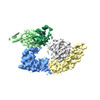
| Title | Cryo-EM structure of a full-length HACE1 dimer | |||||||||
 Map data Map data | ||||||||||
 Sample Sample |
| |||||||||
 Keywords Keywords | E3 / HECT /  ubiquitin / LIGASE ubiquitin / LIGASE | |||||||||
| Function / homology |  Function and homology information Function and homology informationHECT-type E3 ubiquitin transferase / Golgi cisterna membrane / Rac protein signal transduction / Golgi organization / protein K48-linked ubiquitination / regulation of cell migration / small GTPase binding / protein polyubiquitination / ubiquitin-protein transferase activity / ubiquitin protein ligase activity ...HECT-type E3 ubiquitin transferase / Golgi cisterna membrane / Rac protein signal transduction / Golgi organization / protein K48-linked ubiquitination / regulation of cell migration / small GTPase binding / protein polyubiquitination / ubiquitin-protein transferase activity / ubiquitin protein ligase activity / Antigen processing: Ubiquitination & Proteasome degradation / ubiquitin-dependent protein catabolic process / membrane fusion / protein ubiquitination / nuclear body / cell cycle / Golgi membrane / endoplasmic reticulum / nucleus / cytoplasmSimilarity search - Function | |||||||||
| Biological species |  Homo sapiens (human) Homo sapiens (human) | |||||||||
| Method | single particle reconstruction / cryo EM / Resolution: 4.73 Å | |||||||||
 Authors Authors | Duering J / Wolter M / Dienemann C / Lorenz S | |||||||||
| Funding support |  Germany, European Union, 2 items Germany, European Union, 2 items
| |||||||||
 Citation Citation | Journal: Nat Struct Mol Biol / Year: 2024 Title: Structural mechanisms of autoinhibition and substrate recognition by the ubiquitin ligase HACE1. Authors: Jonas Düring / Madita Wolter / Julia J Toplak / Camilo Torres / Olexandr Dybkov / Thornton J Fokkens / Katherine E Bohnsack / Henning Urlaub / Wieland Steinchen / Christian Dienemann / Sonja Lorenz / Abstract: Ubiquitin ligases (E3s) are pivotal specificity determinants in the ubiquitin system by selecting substrates and decorating them with distinct ubiquitin signals. However, structure determination of ...Ubiquitin ligases (E3s) are pivotal specificity determinants in the ubiquitin system by selecting substrates and decorating them with distinct ubiquitin signals. However, structure determination of the underlying, specific E3-substrate complexes has proven challenging owing to their transient nature. In particular, it is incompletely understood how members of the catalytic cysteine-driven class of HECT-type ligases (HECTs) position substrate proteins for modification. Here, we report a cryogenic electron microscopy (cryo-EM) structure of the full-length human HECT HACE1, along with solution-based conformational analyses by small-angle X-ray scattering and hydrogen-deuterium exchange mass spectrometry. Structure-based functional analyses in vitro and in cells reveal that the activity of HACE1 is stringently regulated by dimerization-induced autoinhibition. The inhibition occurs at the first step of the catalytic cycle and is thus substrate-independent. We use mechanism-based chemical crosslinking to reconstitute a complex of activated, monomeric HACE1 with its major substrate, RAC1, determine its structure by cryo-EM and validate the binding mode by solution-based analyses. Our findings explain how HACE1 achieves selectivity in ubiquitinating the active, GTP-loaded state of RAC1 and establish a framework for interpreting mutational alterations of the HACE1-RAC1 interplay in disease. More broadly, this work illuminates central unexplored aspects in the architecture, conformational dynamics, regulation and specificity of full-length HECTs. | |||||||||
| History |
|
- Structure visualization
| Supplemental images |
|---|

- Downloads & links
-EMDB archive
| Map data | emd_17994.map.gz | 97.1 MB | EMDB map data format | |
|---|---|---|---|---|
| Header (meta data) | emd-17994-v30.xmlemd-17994.xml | 16.9 KB 16.9 KB | Display Display | EMDB header |
| FSC (resolution estimation) | emd_17994_fsc.xml | 11.1 KB | Display | FSC data file |
| Images | 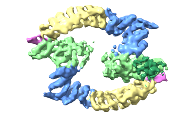 emd_17994.png emd_17994.png | 79.3 KB | ||
| Filedesc metadata | emd-17994.cif.gz | 6.2 KB | ||
| Others | emd_17994_half_map_1.map.gzemd_17994_half_map_2.map.gz | 95.6 MB 95.6 MB | ||
| Archive directory |  http://ftp.pdbj.org/pub/emdb/structures/EMD-17994ftp://ftp.pdbj.org/pub/emdb/structures/EMD-17994 http://ftp.pdbj.org/pub/emdb/structures/EMD-17994ftp://ftp.pdbj.org/pub/emdb/structures/EMD-17994 | HTTPS FTP |
-Related structure data
| Related structure data | 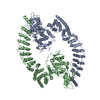 8pwlMC  8q0nC M: atomic model generated by this map C: citing same article ( |
|---|---|
| Similar structure data |
-Links
| EMDB pages | EMDB (EBI/PDBe) / EMDataResource |
|---|
-Map
| File | Download / File: emd_17994.map.gz / Format: CCP4 / Size: 103 MB / Type: IMAGE STORED AS FLOATING POINT NUMBER (4 BYTES) | ||||||||||||||||||||
|---|---|---|---|---|---|---|---|---|---|---|---|---|---|---|---|---|---|---|---|---|---|
| Voxel size | X=Y=Z: 0.834 Å | ||||||||||||||||||||
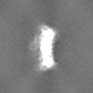
| Density |
| ||||||||||||||||||||
| Symmetry | Space group: 1 | ||||||||||||||||||||
| Details | EMDB XML:
|
-Supplemental data
-Half map: #2
| File | emd_17994_half_map_1.map | ||||||||||||
|---|---|---|---|---|---|---|---|---|---|---|---|---|---|
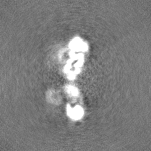
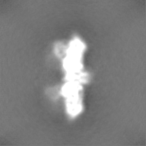
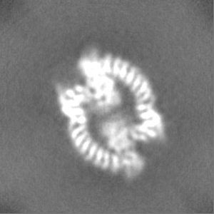
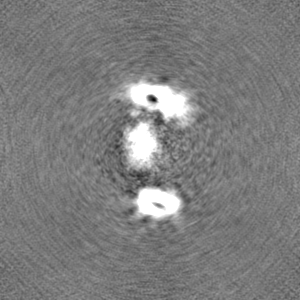
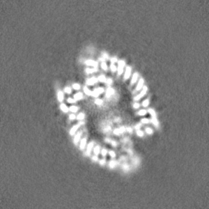
| Projections & Slices |
| ||||||||||||
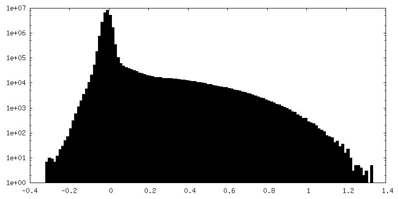
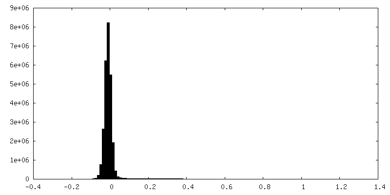
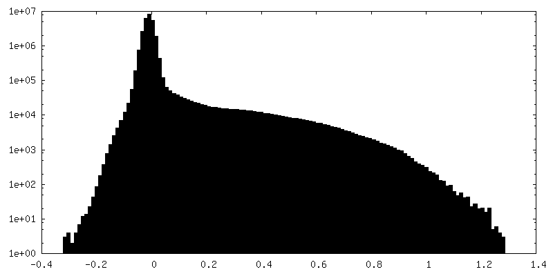
| Density Histograms |
 Z
Z Y
Y X
X


-Half map: #1
| File | emd_17994_half_map_2.map | ||||||||||||
|---|---|---|---|---|---|---|---|---|---|---|---|---|---|
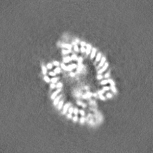
| Projections & Slices |
| ||||||||||||
| Density Histograms |

- Sample components
-Entire : HACE1
| Entire | Name: HACE1 |
|---|---|
| Components |
|
-Supramolecule #1: HACE1
| Supramolecule | Name: HACE1 / type: complex / ID: 1 / Parent: 0 / Macromolecule list: all |
|---|---|
| Source (natural) | Organism: Homo sapiens (human) |
-Macromolecule #1: E3 ubiquitin-protein ligase HACE1
| Macromolecule | Name: E3 ubiquitin-protein ligase HACE1 / type: protein_or_peptide / ID: 1 / Number of copies: 2 / Enantiomer: LEVO / EC number: HECT-type E3 ubiquitin transferase |
|---|---|
| Source (natural) | Organism: Homo sapiens (human) |
| Molecular weight | Theoretical: 102.506719 KDa |
| Recombinant expression | Organism:  Escherichia coli BL21 (bacteria) Escherichia coli BL21 (bacteria) |
| Sequence | String: GMERAMEQLN RLTRSLRRAR TVELPEDNET AVYTLMPMVM ADQHRSVSEL LSNSKFDVNY AFGRVKRSLL HIAANCGSVE CLVLLLKKG ANPNYQDISG CTPLHLAARN GQKKCMSKLL EYSADVNICN NEGLTAIHWL AVNGRTELLH DLVQHVSDVD V EDAMGQTA ...String: GMERAMEQLN RLTRSLRRAR TVELPEDNET AVYTLMPMVM ADQHRSVSEL LSNSKFDVNY AFGRVKRSLL HIAANCGSVE CLVLLLKKG ANPNYQDISG CTPLHLAARN GQKKCMSKLL EYSADVNICN NEGLTAIHWL AVNGRTELLH DLVQHVSDVD V EDAMGQTA LHVACQNGHK TTVQCLLDSG ADINRPNVSG ATPLYFACSH GQRDTAQILL LRGAKYLPDK NGVTPLDLCV QG GYGETCE VLIQYHPRLF QTIIQMTQNE DLRENMLRQV LEHLSQQSES QYLKILTSLA EVATTNGHKL LSLSSNYDAQ MKS LLRIVR MFCHVFRIGP SSPSNGIDMG YNGNKTPRSQ VFKPLELLWH SLDEWLVLIA TELMKNKRDS TEITSILLKQ KGQD QDAAS IPPFEPPGPG SYENLSTGTR ESKPDALAGR QEASADCQDV ISMTANRLSA VIQAFYMCCS CQMPPGMTSP RFIEF VCKH DEVLKCFVNR NPKIIFDHFH FLLECPELMS RFMHIIKAQP FKDRCEWFYE HLHSGQPDSD MVHRPVNEND ILLVHR DSI FRSSCEVVSK ANCAKLKQGI AVRFHGEEGM GQGVVREWFD ILSNEIVNPD YALFTQSADG TTFQPNSNSY VNPDHLN YF RFAGQILGLA LNHRQLVNIY FTRSFYKHIL GIPVNYQDVA SIDPEYAKNL QWILDNDISD LGLELTFSVE TDVFGAME E VPLKPGGGSI LVTQNNKAEY VQLVTELRMT RAIQPQINAF LQGFHMFIPP SLIQLFDEYE LELLLSGMPE IDVSDWIKN TEYTSGYERE DPVIQWFWEV VEDITQEERV LLLQFVTGSS RVPHGGFANI MGGSGLQNFT IAAVPYTPNL LPTSSTCINM LKLPEYPSK EILKDRLLVA LHCGSYGYTM A UniProtKB: E3 ubiquitin-protein ligase HACE1 |
-Experimental details
-Structure determination
| Method | cryo EM |
|---|---|
 Processing Processing | single particle reconstruction |
| Aggregation state | particle |
-Sample preparation
| Concentration | 0.7 mg/mL | ||||||||||||
|---|---|---|---|---|---|---|---|---|---|---|---|---|---|
| Buffer | pH: 8 Component:
| ||||||||||||
| Grid | Model: Quantifoil R2/1 / Material: COPPER / Mesh: 400 / Pretreatment - Type: GLOW DISCHARGE | ||||||||||||
| Vitrification | Cryogen name: ETHANE / Chamber humidity: 95 % / Chamber temperature: 277 K |
- Electron microscopy
| Microscope | FEI TITAN KRIOS |
|---|---|
| Electron beam | Acceleration voltage: 300 kV / Electron source: FIELD EMISSION GUN |
| Electron optics | Illumination mode: FLOOD BEAM / Imaging mode: BRIGHT FIELDBright-field microscopy / Cs: 2.7 mm / Nominal defocus max: 3.2 µm / Nominal defocus min: 0.3 µm / Nominal magnification: 105000 |
| Image recording | Film or detector model: GATAN K3 (6k x 4k) / Number grids imaged: 2 / Number real images: 29748 / Average electron dose: 40.0 e/Å2 |
| Experimental equipment |  Model: Titan Krios / Image courtesy: FEI Company |
-Image processing
| Particle selection | Number selected: 3639240 |
|---|---|
| Startup model | Type of model: NONE |
| Initial angle assignment | Type: MAXIMUM LIKELIHOOD / Software - Name: cryoSPARC (ver. v4) |
| Final angle assignment | Type: MAXIMUM LIKELIHOOD / Software - Name: cryoSPARC (ver. v4) |
| Final reconstruction | Applied symmetry - Point group: C1 (asymmetric) / Resolution.type: BY AUTHOR / Resolution: 4.73 Å / Resolution method: FSC 0.143 CUT-OFF / Software - Name: cryoSPARC (ver. v4) / Number images used: 118791 |
| FSC plot (resolution estimation) | 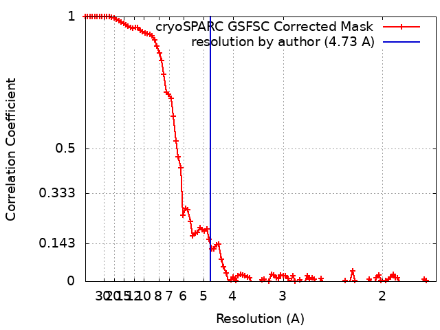 |
-Atomic model buiding 1
| Initial model | Chain - Source name: AlphaFold / Chain - Initial model type: in silico model |
|---|---|
| Refinement | Space: REAL / Protocol: RIGID BODY FIT |
| Output model | PDB-8pwl: |
