 Movie
Movie Controller


+ Open data

- Basic information
| Entry |  | |||||||||
|---|---|---|---|---|---|---|---|---|---|---|
| Title | Structure of divisome complex FtsWIQLB | |||||||||
 Map data Map data | ||||||||||
 Sample Sample |
| |||||||||
 Keywords Keywords | FtsWIQLB /  Gram-negative bacteria / membrane protein / divisome / CELL CYCLE Gram-negative bacteria / membrane protein / divisome / CELL CYCLE | |||||||||
| Function / homology |  Function and homology information Function and homology informationcell septum assembly / lipid-linked peptidoglycan transporter activity / peptidoglycan glycosyltransferase / peptidoglycan glycosyltransferase activity / cell septum / serine-type D-Ala-D-Ala carboxypeptidase / FtsZ-dependent cytokinesis / serine-type D-Ala-D-Ala carboxypeptidase activity / division septum assembly / cell division site ...cell septum assembly / lipid-linked peptidoglycan transporter activity / peptidoglycan glycosyltransferase / peptidoglycan glycosyltransferase activity / cell septum / serine-type D-Ala-D-Ala carboxypeptidase / FtsZ-dependent cytokinesis / serine-type D-Ala-D-Ala carboxypeptidase activity / division septum assembly / cell division site / penicillin binding / peptidoglycan biosynthetic process / cell wall organization / regulation of cell shape / cell division / proteolysis / plasma membraneSimilarity search - Function | |||||||||
| Biological species |  Escherichia coli K-12 (bacteria) Escherichia coli K-12 (bacteria) | |||||||||
| Method | single particle reconstruction / cryo EM / Resolution: 3.3 Å | |||||||||
 Authors Authors | Yang L / Chang S / Tang D / Dong H / Xie T / Luo B / Lu G / Zhu X / Wei X / Dong C ...Yang L / Chang S / Tang D / Dong H / Xie T / Luo B / Lu G / Zhu X / Wei X / Dong C / Zhou R / Zhang X / Tang X | |||||||||
| Funding support |  China, 1 items China, 1 items
| |||||||||
 Citation Citation | Journal: Cell Discov / Year: 2024 Title: Structural insights into the activation of the divisome complex FtsWIQLB. Authors: Lili Yang / Yujiao Chen / Shenghai Chang / Chongrong Shen / Xin Wang / Changbin Zhang / Zhibo Zhang / Bi-Sen Ding / Zhaoming Su / Haohao Dong / Xiaodi Tang / | |||||||||
| History |
|
- Structure visualization
| Supplemental images |
|---|

- Downloads & links
-EMDB archive
| Map data | emd_17356.map.gz | 97.2 MB | EMDB map data format | |
|---|---|---|---|---|
| Header (meta data) | emd-17356-v30.xmlemd-17356.xml | 19.2 KB 19.2 KB | Display Display | EMDB header |
| FSC (resolution estimation) | emd_17356_fsc.xml | 9.9 KB | Display | FSC data file |
| Images | 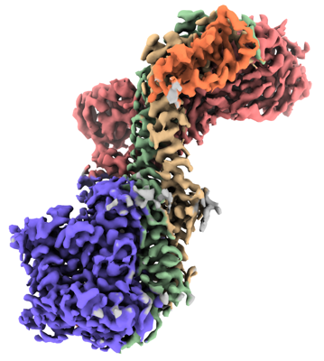 emd_17356.png emd_17356.png | 135.6 KB | ||
| Filedesc metadata | emd-17356.cif.gz | 6.3 KB | ||
| Others | emd_17356_half_map_1.map.gzemd_17356_half_map_2.map.gz | 95.5 MB 95.5 MB | ||
| Archive directory |  http://ftp.pdbj.org/pub/emdb/structures/EMD-17356ftp://ftp.pdbj.org/pub/emdb/structures/EMD-17356 http://ftp.pdbj.org/pub/emdb/structures/EMD-17356ftp://ftp.pdbj.org/pub/emdb/structures/EMD-17356 | HTTPS FTP |
-Related structure data
| Related structure data |  8p1uMC M: atomic model generated by this map C: citing same article ( |
|---|---|
| Similar structure data |
-Links
| EMDB pages | EMDB (EBI/PDBe) / EMDataResource |
|---|---|
| Related items in Molecule of the Month |

-Map
| File | Download / File: emd_17356.map.gz / Format: CCP4 / Size: 103 MB / Type: IMAGE STORED AS FLOATING POINT NUMBER (4 BYTES) | ||||||||||||||||||||
|---|---|---|---|---|---|---|---|---|---|---|---|---|---|---|---|---|---|---|---|---|---|
| Voxel size | X=Y=Z: 0.93 Å | ||||||||||||||||||||
| Density |
| ||||||||||||||||||||
| Symmetry | Space group: 1 | ||||||||||||||||||||
| Details | EMDB XML:
|
-Supplemental data
-Half map: #2
| File | emd_17356_half_map_1.map | ||||||||||||
|---|---|---|---|---|---|---|---|---|---|---|---|---|---|
| Projections & Slices |
| ||||||||||||
| Density Histograms |
 Z
Z Y
Y X
X


-Half map: #1
| File | emd_17356_half_map_2.map | ||||||||||||
|---|---|---|---|---|---|---|---|---|---|---|---|---|---|
| Projections & Slices |
| ||||||||||||
| Density Histograms |


- Sample components
-Entire : divisome complex FtsWIQLB
| Entire | Name: divisome complex FtsWIQLB |
|---|---|
| Components |
|
-Supramolecule #1: divisome complex FtsWIQLB
| Supramolecule | Name: divisome complex FtsWIQLB / type: complex / ID: 1 / Parent: 0 / Macromolecule list: all |
|---|---|
| Source (natural) | Organism: Escherichia coli K-12 (bacteria) |
-Macromolecule #1: Cell division protein FtsL
| Macromolecule | Name: Cell division protein FtsL / type: protein_or_peptide / ID: 1 / Number of copies: 1 / Enantiomer: LEVO |
|---|---|
| Source (natural) | Organism: Escherichia coli K-12 (bacteria) |
| Molecular weight | Theoretical: 11.150034 KDa |
| Recombinant expression | Organism: Escherichia coli K-12 (bacteria) |
| Sequence | String: MSRLFVKRLP TGSFLMLLLY IGLLLSAIAV AYSTYWNRQL LNSLYSELSV RDKAQAEWGR LILEQSTWTA HSRIESLAVE QLRMRVPDP AEVRMVAP UniProtKB: Cell division protein FtsL |
-Macromolecule #2: Probable peptidoglycan glycosyltransferase FtsW
| Macromolecule | Name: Probable peptidoglycan glycosyltransferase FtsW / type: protein_or_peptide / ID: 2 / Number of copies: 1 / Enantiomer: LEVO / EC number: peptidoglycan glycosyltransferase |
|---|---|
| Source (natural) | Organism: Escherichia coli K-12 (bacteria) |
| Molecular weight | Theoretical: 43.793629 KDa |
| Recombinant expression | Organism: Escherichia coli K-12 (bacteria) |
| Sequence | String: MLSVLRPFPS PLLSRHGIDL DFPLLAGCLA LLGLGLVMVT SASSEVAAAQ SGNPLYFSVR HLIYLVIGLI SCGLTMMVPM ATWQRWGWK LLLVAFGLLV LVITPGIGRE VNGSMRWIGF GLFNIQPSEI AKVCVVIFMA GYLIRRQQEV RESWMGFFKP F VVLLPMAG ...String: MLSVLRPFPS PLLSRHGIDL DFPLLAGCLA LLGLGLVMVT SASSEVAAAQ SGNPLYFSVR HLIYLVIGLI SCGLTMMVPM ATWQRWGWK LLLVAFGLLV LVITPGIGRE VNGSMRWIGF GLFNIQPSEI AKVCVVIFMA GYLIRRQQEV RESWMGFFKP F VVLLPMAG LLLREPDFGA TVVMMGAAAA MLFLGGVGLF RFGLMVLLAV GAVVLLIQTQ PYRMARLTNF TDPWADQFGA GY QLSQALI AFGRGGWLGM GLGNSIQKQF YLPEAHTDFV FAVLAEELGI VGALATVALF VFVSLRALYI GIWAEQAKQF FSA YVAYGL AFLWIGQFLI NIGVNVGLLP TKGLTLPFLS YGGSSLVICC ACLGMLLRIE WERRTHLGSE EYEFNEEDFA DER UniProtKB: Probable peptidoglycan glycosyltransferase FtsW |
-Macromolecule #3: Peptidoglycan D,D-transpeptidase FtsI
| Macromolecule | Name: Peptidoglycan D,D-transpeptidase FtsI / type: protein_or_peptide / ID: 3 / Number of copies: 1 / Enantiomer: LEVO / EC number: serine-type D-Ala-D-Ala carboxypeptidase |
|---|---|
| Source (natural) | Organism: Escherichia coli K-12 (bacteria) |
| Molecular weight | Theoretical: 62.933082 KDa |
| Recombinant expression | Organism: Escherichia coli K-12 (bacteria) |
| Sequence | String: MKLNYFQGAL YPWRFCVIVG LLLAMVGAIV WRIVDLHVID HDFLKGQGDA RSVRHIAIPA HRGLITDRNG EPLAVSTPVT TLWANPKEL MTAKERWPQL AAALGQDTKL FADRIEQNAE REFIYLVRGL TPEQGEGVIA LKVPGVYSIE EFRRFYPAGE V VAHAVGFT ...String: MKLNYFQGAL YPWRFCVIVG LLLAMVGAIV WRIVDLHVID HDFLKGQGDA RSVRHIAIPA HRGLITDRNG EPLAVSTPVT TLWANPKEL MTAKERWPQL AAALGQDTKL FADRIEQNAE REFIYLVRGL TPEQGEGVIA LKVPGVYSIE EFRRFYPAGE V VAHAVGFT DVDDRGREGI ELAFDEWLAG VPGKRQVLKD RRGRVIKDVQ VTKNAKPGKT LALSIDLRLQ YLAHRELRNA LL ENGAKAG SLVIMDVKTG EILAMTNQPT YNPNNRRNLQ PAAMRNRAMI DVFEPGSTVK PFSMSAALAS GRWKPSDIVD VYP GTLQIG RYTIRDVSRN SRQLDLTGIL IKSSNVGISK IAFDIGAESI YSVMQQVGLG QDTGLGFPGE RVGNLPNHRK WPKA ETATL AYGYGLSVTA IQLAHAYAAL ANDGKSVPLS MTRVDRVPDG VQVISPEVAS TVQGMLQQVV EAQGGVFRAQ VPGYH AAGK SGTARKVSVG TKGYRENAYR SLFAGFAPAT DPRIAMVVVI DEPSKAGYFG GLVSAPVFSK VMAGALRLMN VPPDNL PTA TEQQQVNAAP AKGGRG UniProtKB: Peptidoglycan D,D-transpeptidase FtsI |
-Macromolecule #4: Cell division protein FtsB
| Macromolecule | Name: Cell division protein FtsB / type: protein_or_peptide / ID: 4 / Number of copies: 1 / Enantiomer: LEVO |
|---|---|
| Source (natural) | Organism: Escherichia coli K-12 (bacteria) |
| Molecular weight | Theoretical: 10.890521 KDa |
| Recombinant expression | Organism: Escherichia coli K-12 (bacteria) |
| Sequence | String: MRLRSPYWLF VVLILALAGL QYRLWVGDGS LAQVRDLQKQ IADQHGENER LLERNRILEA EVAELKKGTE TVEERARHEL GMVKDGETL YQLAK UniProtKB: Cell division protein FtsB |
-Macromolecule #5: Cell division protein FtsQ
| Macromolecule | Name: Cell division protein FtsQ / type: protein_or_peptide / ID: 5 / Number of copies: 1 / Enantiomer: LEVO |
|---|---|
| Source (natural) | Organism: Escherichia coli K-12 (bacteria) |
| Molecular weight | Theoretical: 32.290223 KDa |
| Recombinant expression | Organism: Escherichia coli K-12 (bacteria) |
| Sequence | String: MNGVLLRHQQ PGGLGRAPRK PMPRGASRLV AKEPLSVRLP KADFSFLKYL AWPLLLAVLG YGAYRGAEYI LPYADRPIAK VSVEGDLSY ISQRAVQQRI SPYLAASFFT IDLAGMRGQL EQMPWIAHAE VRRVWPDQVV IRLDEQLPIA RWGDEALLNN Q GQAFTPKE ...String: MNGVLLRHQQ PGGLGRAPRK PMPRGASRLV AKEPLSVRLP KADFSFLKYL AWPLLLAVLG YGAYRGAEYI LPYADRPIAK VSVEGDLSY ISQRAVQQRI SPYLAASFFT IDLAGMRGQL EQMPWIAHAE VRRVWPDQVV IRLDEQLPIA RWGDEALLNN Q GQAFTPKE LANYEHLPRL HGPQRAQQQV MQQYQLLSQL LRPLGFSIAR LEMSDRGGWA LTTAQGVEIQ IGRDHVVDKI RR FVSIYDK ALKDQISNIA RIDLRYPNGL AVAWREPVTP ATVATASAVQ UniProtKB: Cell division protein FtsQ |
-Experimental details
-Structure determination
| Method | cryo EM |
|---|---|
 Processing Processing | single particle reconstruction |
| Aggregation state | particle |
-Sample preparation
| Buffer | pH: 7.8 |
|---|---|
| Vitrification | Cryogen name: ETHANE |
- Electron microscopy
| Microscope | FEI TITAN KRIOS |
|---|---|
| Electron beam | Acceleration voltage: 300 kV / Electron source: FIELD EMISSION GUN |
| Electron optics | Calibrated defocus max: 2.0 µm / Calibrated defocus min: 1.0 µm / Illumination mode: FLOOD BEAM / Imaging mode: BRIGHT FIELDBright-field microscopy / Nominal defocus max: 2.0 µm / Nominal defocus min: 1.0 µm |
| Image recording | Film or detector model: FEI FALCON IV (4k x 4k) / Average electron dose: 50.0 e/Å2 |
| Experimental equipment |  Model: Titan Krios / Image courtesy: FEI Company |
-Image processing
| Startup model | Type of model: NONE |
|---|---|
| Initial angle assignment | Type: MAXIMUM LIKELIHOOD |
| Final angle assignment | Type: MAXIMUM LIKELIHOOD |
| Final reconstruction | Resolution.type: BY AUTHOR / Resolution: 3.3 Å / Resolution method: FSC 0.143 CUT-OFF / Number images used: 340758 |
| FSC plot (resolution estimation) |  |
