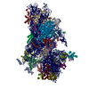
Translation initiation complex formation / Formation of the ternary complex, and subsequently, the 43S complex / Ribosomal scanning and start codon recognition / Major pathway of rRNA processing in the nucleolus and cytosol / GTP hydrolysis and joining of the 60S ribosomal subunit / L13a-mediated translational silencing of Ceruloplasmin expression / SRP-dependent cotranslational protein targeting to membrane / Formation of a pool of free 40S subunits / Nonsense Mediated Decay (NMD) independent of the Exon Junction Complex (EJC) / Nonsense Mediated Decay (NMD) enhanced by the Exon Junction Complex (EJC) ...Translation initiation complex formation / Formation of the ternary complex, and subsequently, the 43S complex / Ribosomal scanning and start codon recognition / Major pathway of rRNA processing in the nucleolus and cytosol / GTP hydrolysis and joining of the 60S ribosomal subunit / L13a-mediated translational silencing of Ceruloplasmin expression / SRP-dependent cotranslational protein targeting to membrane / Formation of a pool of free 40S subunits / Nonsense Mediated Decay (NMD) independent of the Exon Junction Complex (EJC) / Nonsense Mediated Decay (NMD) enhanced by the Exon Junction Complex (EJC) / ribosomal subunit / endonucleolytic cleavage to generate mature 3'-end of SSU-rRNA from (SSU-rRNA, 5.8S rRNA, LSU-rRNA) / endonucleolytic cleavage in ITS1 to separate SSU-rRNA from 5.8S rRNA and LSU-rRNA from tricistronic rRNA transcript (SSU-rRNA, 5.8S rRNA, LSU-rRNA) / rough endoplasmic reticulum / regulation of translational fidelity / translation initiation factor activity / DNA-(apurinic or apyrimidinic site) lyase / small-subunit processome / ribosomal small subunit biogenesis / cytoplasmic stress granule / cytosolic small ribosomal subunit / cytoplasmic translation / cell differentiation / ribosome / structural constituent of ribosome / ribonucleoprotein complex / translation / mRNA binding / synapse / nucleolus / ATP hydrolysis activity / RNA binding / ATP binding / cytoplasm Similarity search - Function
RLI, domain 1 / RLI1 / RNase L inhibitor RLI-like, possible metal-binding domain / Possible Fer4-like domain in RNase L inhibitor, RLI / Translation initiation factor 1A (eIF-1A), conserved site / Eukaryotic initiation factor 1A signature. / eukaryotic translation initiation factor 1A / Translation initiation factor 1A (eIF-1A) / IF2a, S1-like domain / Translation initiation factor 2, alpha subunit ...RLI, domain 1 / RLI1 / RNase L inhibitor RLI-like, possible metal-binding domain / Possible Fer4-like domain in RNase L inhibitor, RLI / Translation initiation factor 1A (eIF-1A), conserved site / Eukaryotic initiation factor 1A signature. / eukaryotic translation initiation factor 1A / Translation initiation factor 1A (eIF-1A) / IF2a, S1-like domain / Translation initiation factor 2, alpha subunit / Translation initiation factor 2, alpha subunit, middle domain superfamily / Translation initiation factor 2, alpha subunit, C-terminal / Eukaryotic translation initiation factor 2 alpha subunit / RNA-binding domain, S1, IF1 type / Translation initiation factor 1A / IF-1 / S1 domain IF1 type profile. / 4Fe-4S binding domain / Ribosomal protein S21e, conserved site / Ribosomal protein S26e signature. / S1 domain profile. / Ribosomal protein S21e / Ribosomal protein S21e superfamily / Ribosomal protein S21e / Ribosomal protein S21e signature. / Ribosomal protein S3Ae, conserved site / Ribosomal protein S12e signature. / 40S ribosomal protein S1/3, eukaryotes / Ribosomal protein S6, eukaryotic / Ribosomal protein S19e signature. / 40S Ribosomal protein S10 / Ribosomal protein S6/S6e/A/B/2, conserved site / Plectin/S10, N-terminal / Ribosomal protein S3Ae / Ribosomal S3Ae family / Plectin/S10 domain / Ribosomal protein S5/S7, eukaryotic/archaeal / Ribosomal protein S6e / Ribosomal protein S6e / Ribosomal S3Ae family / Ribosomal protein S17e signature. / Ribosomal protein S7e signature. / Ribosomal protein S6e / Ribosomal protein S3Ae signature. / Ribosomal protein S27e signature. / Ribosomal protein S4e signature. / Ribosomal protein S8e signature. / Ribosomal S24e conserved site / Ribosomal protein S24e signature. / Ribosomal protein S24e / Ribosomal protein S24e / Ribosomal protein S6e signature. / Ribosomal protein S28e signature. / Ribosomal protein S1-like RNA-binding domain / S1 RNA binding domain / S1 domain / 4Fe-4S ferredoxin, iron-sulphur binding, conserved site / 4Fe-4S ferredoxin-type iron-sulfur binding region signature. / 4Fe-4S ferredoxin-type iron-sulfur binding domain profile. / 4Fe-4S ferredoxin-type, iron-sulphur binding domain / ABC transporter-like, conserved site / ABC transporters family signature. / ABC transporter / ABC transporter-like, ATP-binding domain / ATP-binding cassette, ABC transporter-type domain profile. / Ribosomal protein S2 signature 2. / Ubiquitin domain signature. / Ribosomal protein L30 signature. / Ribosomal protein S3 signature. / Ribosomal protein S10 signature. / Ribosomal protein S14 signature. / Ribosomal protein S7, conserved site / Ribosomal protein S2 signature 1. / Type-2 KH domain profile. / Ribosomal protein S19 signature. / Ribosomal protein S7 signature. / Ribosomal protein S17 signature. / Ribosomal protein S13 signature. / Ribosomal protein S5/S7 / Ribosomal protein S7 domain / Ribosomal protein S7 domain superfamily / Ribosomal protein S13 family profile. / Ribosomal protein S8 signature. / Ribosomal protein S4 signature. / Ribosomal protein S15 signature. / Ribosomal protein S7p/S5e / S4 RNA-binding domain profile. / Ribosomal protein S11 signature. / Ubiquitin domain profile. / Ribosomal protein S9 signature. / Ribosomal protein S12 signature. / Ribosomal protein L23/L15e core domain superfamily / Trp-Asp (WD) repeats signature. / Trp-Asp (WD) repeats profile. / Trp-Asp (WD) repeats circular profile. / Winged helix-like DNA-binding domain superfamily / ATPases associated with a variety of cellular activities / AAA+ ATPase domain / Nucleic acid-binding, OB-fold / P-loop containing nucleoside triphosphate hydrolase Similarity search - Domain/homology
Small ribosomal subunit protein eS32 / 40S ribosomal protein S6 / Small ribosomal subunit protein uS4 / Small ribosomal subunit protein eS12 / ATP binding cassette subfamily E member 1 / Small ribosomal subunit protein uS9 / Small ribosomal subunit protein uS10 / Small ribosomal subunit protein RACK1 / Ubiquitin-ribosomal protein eS31 fusion protein / Small ribosomal subunit protein uS15 ...Small ribosomal subunit protein eS32 / 40S ribosomal protein S6 / Small ribosomal subunit protein uS4 / Small ribosomal subunit protein eS12 / ATP binding cassette subfamily E member 1 / Small ribosomal subunit protein uS9 / Small ribosomal subunit protein uS10 / Small ribosomal subunit protein RACK1 / Ubiquitin-ribosomal protein eS31 fusion protein / Small ribosomal subunit protein uS15 / Small ribosomal subunit protein eS7 / Eukaryotic translation initiation factor 4C / Small ribosomal subunit protein uS12 / Small ribosomal subunit protein uS11 / Eukaryotic translation initiation factor 2 subunit 1 / Ubiquitin-like FUBI-ribosomal protein eS30 fusion protein / Small ribosomal subunit protein eS25 / Small ribosomal subunit protein eS26 / Small ribosomal subunit protein uS8 / Small ribosomal subunit protein eS28 / Small ribosomal subunit protein eS8 / Small ribosomal subunit protein eS4 / Small ribosomal subunit protein uS2 / Small ribosomal subunit protein eS19 / Small ribosomal subunit protein eS1 / Small ribosomal subunit protein uS3 / Small ribosomal subunit protein uS13 / Plectin/eS10 N-terminal domain-containing protein / Small ribosomal subunit protein uS17 / 40S ribosomal protein S24 / Small ribosomal subunit protein eS17 / Large ribosomal subunit protein uL30 / Small ribosomal subunit protein eS27 / Small ribosomal subunit protein uS19 / Small ribosomal subunit protein uS14 / Small ribosomal subunit protein eS21 / Ribosomal protein S5 Similarity search - Component
Biological species
Oryctolagus cuniculus (rabbit)
Method
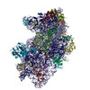
single particle reconstruction / cryo EM / Resolution: 3.04 Å
Journal: Mol Cell / Year: 2024 Title: N-methyladenosine in 5' UTR does not promote translation initiation. Authors: Ewelina Guca / Rodrigo Alarcon / Michael Z Palo / Leonardo Santos / Santiago Alonso-Gil / Marcos Davyt / Leonardo H F de Lima / Fanny Boissier / Sarada Das / Bojan Zagrovic / Joseph D ...Authors: Ewelina Guca / Rodrigo Alarcon / Michael Z Palo / Leonardo Santos / Santiago Alonso-Gil / Marcos Davyt / Leonardo H F de Lima / Fanny Boissier / Sarada Das / Bojan Zagrovic / Joseph D Puglisi / Yaser Hashem / Zoya Ignatova / Abstract: The most abundant N-methyladenosine (mA) modification on mRNAs is installed non-stoichiometrically across transcripts, with 5' untranslated regions (5' UTRs) being the least conductive. 5' UTRs are ...The most abundant N-methyladenosine (mA) modification on mRNAs is installed non-stoichiometrically across transcripts, with 5' untranslated regions (5' UTRs) being the least conductive. 5' UTRs are essential for translation initiation, yet the molecular mechanisms orchestrated by mA remain poorly understood. Here, we combined structural, biochemical, and single-molecule approaches and show that at the most common position, a single mA does not affect translation yields, the kinetics of translation initiation complex assembly, or start codon recognition both under permissive growth and following exposure to oxidative stress. Cryoelectron microscopy (cryo-EM) structures of the late preinitiation complex reveal that mA purine ring established stacking interactions with an arginine side chain of the initiation factor eIF2α, although with only a marginal energy contribution, as estimated computationally. These findings provide molecular insights into mA interactions with the initiation complex and suggest that the subtle stabilization is unlikely to affect the translation dynamics under homeostatic conditions or stress.
In the structure databanks used in Yorodumi, some data are registered as the other names, "COVID-19 virus" and "2019-nCoV". Here are the details of the virus and the list of structure data.
Jan 31, 2019. EMDB accession codes are about to change! (news from PDBe EMDB page)
EMDB accession codes are about to change! (news from PDBe EMDB page)
The allocation of 4 digits for EMDB accession codes will soon come to an end. Whilst these codes will remain in use, new EMDB accession codes will include an additional digit and will expand incrementally as the available range of codes is exhausted. The current 4-digit format prefixed with “EMD-” (i.e. EMD-XXXX) will advance to a 5-digit format (i.e. EMD-XXXXX), and so on. It is currently estimated that the 4-digit codes will be depleted around Spring 2019, at which point the 5-digit format will come into force.
The EM Navigator/Yorodumi systems omit the EMD- prefix.
Related info.:Q: What is EMD? / ID/Accession-code notation in Yorodumi/EM Navigator
Yorodumi is a browser for structure data from EMDB, PDB, SASBDB, etc.
This page is also the successor to EM Navigator detail page, and also detail information page/front-end page for Omokage search.
The word "yorodu" (or yorozu) is an old Japanese word meaning "ten thousand". "mi" (miru) is to see.
Related info.:EMDB / PDB / SASBDB / Comparison of 3 databanks / Yorodumi Search / Aug 31, 2016. New EM Navigator & Yorodumi / Yorodumi Papers / Jmol/JSmol / Function and homology information / Changes in new EM Navigator and Yorodumi
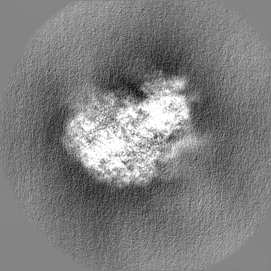
 Movie
Movie Controller


 Open data
 Basic information
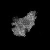
 Map data
 Sample
 Keywords
 translation initiation /
  Function and homology information
 Authors
 Citation


 Structure visualization
 Downloads & links
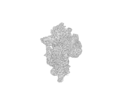 emd_17329.png
 http://ftp.pdbj.org/pub/emdb/structures/EMD-17329
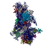


 Z (Sec.)
 Y (Row.)
 X (Col.)


 Sample components
 Processing
 Electron microscopy